当前位置:
X-MOL 学术
›
Microsc. Res. Tech.
›
论文详情
Our official English website, www.x-mol.net, welcomes your feedback! (Note: you will need to create a separate account there.)
Scanning electron microscopy comparison of the resin-dentin interface using different specimen preparation methods.
Microscopy Research and Technique ( IF 2.5 ) Pub Date : 2020-04-11 , DOI: 10.1002/jemt.23488 Marina G Augusto 1 , Debora C B Dantas 1 , Guilherme S de Andrade 2 , Amanda G N Matuda 1 , Stephanie R Lopes 1 , Daphne C Barcellos 1 , Cesar R Pucci 1
Microscopy Research and Technique ( IF 2.5 ) Pub Date : 2020-04-11 , DOI: 10.1002/jemt.23488 Marina G Augusto 1 , Debora C B Dantas 1 , Guilherme S de Andrade 2 , Amanda G N Matuda 1 , Stephanie R Lopes 1 , Daphne C Barcellos 1 , Cesar R Pucci 1
Affiliation
|
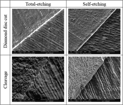
Microscopy has been widely used to complement the data of studies related to dentin bonding; however, different specimen preparation methods may influence the analysis. Aiming to contribute to the reported scenario, this study evaluated the effect of two specimen‐sectioning methods (cleavage and diamond disk cut) on the quality of the scanning electron microscopy (SEM) images. Four crowns of human molars were selected and had an area of approximately 6 mm2 of dentin exposed. They were then divided into two groups according to the universal adhesive application: total and self‐etching modes. Then, composite blocks were built up and the specimens were stored in deionized water to allow the postcuring. The specimens were further divided according to the sectioning method: cleavage or diamond disk cut. Four specimens were obtained from each tooth. They were desiccated, placed on aluminum stubs, sputter‐coated with gold, and observed in a scanning electron microscope, with ×2000 of magnification. The quality of the SEM images were evaluated by two calibrated examiners and classified into four scores (1–4). Mann–Whitney test (p < .05) showed that the diamond disk provided significantly higher scores than cleavage, whereas no significant difference was observed when comparing the total‐etching and self‐etching modes of application. The diamond disk cut method is preferable to the cleavage method to ensure the quality of the SEM analysis in studies involving the resin–dentin interface.
中文翻译:

使用不同样品制备方法的树脂-牙本质界面的扫描电子显微镜比较。
显微镜已被广泛用于补充与牙本质粘合有关的研究数据。但是,不同的样品制备方法可能会影响分析。为了对所报告的情况做出贡献,本研究评估了两种标本切片方法(切割和钻石盘切割)对扫描电子显微镜(SEM)图像质量的影响。选择四颗人类臼齿的牙冠,其面积约为6 mm 2的牙本质暴露。然后根据通用胶粘剂的应用将它们分为两组:整体和自蚀刻模式。然后,建立复合块,并将样品存储在去离子水中以进行后固化。根据切片方法将标本进一步分割:劈开或金刚石盘切割。从每个牙齿获得四个样本。将它们干燥,放在铝制的短棒上,用金喷镀,然后在扫描电子显微镜中观察,放大倍数为2000。SEM图像的质量由两名经过校准的检查员进行评估,并分为四个分数(1-4)。曼惠特尼检验(p <.05)表明,金刚石盘的得分明显高于劈裂,而比较总蚀刻和自蚀刻的应用模式时,则观察不到明显差异。在涉及树脂-牙本质界面的研究中,金刚石圆盘切割法比裂解法更好,以确保SEM分析的质量。
更新日期:2020-04-11
中文翻译:
使用不同样品制备方法的树脂-牙本质界面的扫描电子显微镜比较。
显微镜已被广泛用于补充与牙本质粘合有关的研究数据。但是,不同的样品制备方法可能会影响分析。为了对所报告的情况做出贡献,本研究评估了两种标本切片方法(切割和钻石盘切割)对扫描电子显微镜(SEM)图像质量的影响。选择四颗人类臼齿的牙冠,其面积约为6 mm 2的牙本质暴露。然后根据通用胶粘剂的应用将它们分为两组:整体和自蚀刻模式。然后,建立复合块,并将样品存储在去离子水中以进行后固化。根据切片方法将标本进一步分割:劈开或金刚石盘切割。从每个牙齿获得四个样本。将它们干燥,放在铝制的短棒上,用金喷镀,然后在扫描电子显微镜中观察,放大倍数为2000。SEM图像的质量由两名经过校准的检查员进行评估,并分为四个分数(1-4)。曼惠特尼检验(p <.05)表明,金刚石盘的得分明显高于劈裂,而比较总蚀刻和自蚀刻的应用模式时,则观察不到明显差异。在涉及树脂-牙本质界面的研究中,金刚石圆盘切割法比裂解法更好,以确保SEM分析的质量。


 京公网安备 11010802027423号