Our official English website, www.x-mol.net, welcomes your feedback! (Note: you will need to create a separate account there.)
Microglia senescence occurs in both substantia nigra and ventral tegmental area.
Glia ( IF 6.2 ) Pub Date : 2020-04-10 , DOI: 10.1002/glia.23834 Fatemeh Shaerzadeh 1 , Leah Phan 1 , Douglas Miller 1 , Maxwell Dacquel 1 , William Hachmeister 1 , Carissa Hansen 1 , Alexandra Bechtle 1 , Duan Tu 2 , Maia Martcheva 2 , Thomas C Foster 1 , Ashok Kumar 1 , Wolfgang J Streit 1 , Habibeh Khoshbouei 1
Glia ( IF 6.2 ) Pub Date : 2020-04-10 , DOI: 10.1002/glia.23834 Fatemeh Shaerzadeh 1 , Leah Phan 1 , Douglas Miller 1 , Maxwell Dacquel 1 , William Hachmeister 1 , Carissa Hansen 1 , Alexandra Bechtle 1 , Duan Tu 2 , Maia Martcheva 2 , Thomas C Foster 1 , Ashok Kumar 1 , Wolfgang J Streit 1 , Habibeh Khoshbouei 1
Affiliation
|
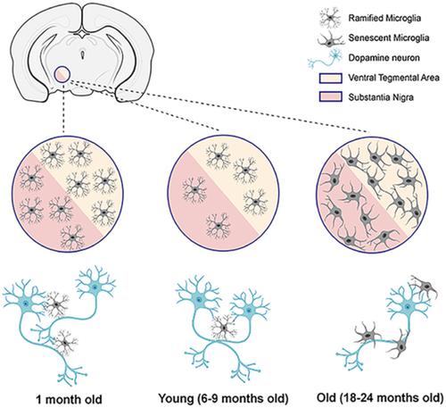
During aging humans lose midbrain dopamine neurons, but not all dopamine regions exhibit vulnerability to neurodegeneration. Microglia maintain tissue homeostasis and neuronal support, but microglia become senescent and likely lose some of their functional abilities. Since aging is the greatest risk factor for Parkinson's disease, we hypothesized that aging‐related changes in microglia and neurons occur in the vulnerable substantia nigra pars compacta (SNc) but not the ventral tegmental area (VTA). We conducted stereological analyses to enumerate microglia and dopaminergic neurons in the SNc and VTA of 1‐, 6‐, 9‐, 18‐, and 24‐month‐old C57BL/J6 mice using sections double‐stained with tyrosine hydroxylase (TH) and Iba1. Both brain regions show an increase in microglia with aging, whereas numbers of TH+ cells show no significant change after 9 months of age in SNc and 6 months in VTA. Morphometric analyses reveal reduced microglial complexity and projection area while cell body size increases with aging. Contact sites between microglia and dopaminergic neurons in both regions increase with aging, suggesting increased microglial support/surveillance of dopamine neurons. To assess neurotrophin expression in dopaminergic neurons, BDNF and TH mRNA were quantified. Results show that the ratio of BDNF to TH decreases in the SNc, but not the VTA. Gait analysis indicates subtle, aging‐dependent changes in gait indices. In conclusion, increases in microglial cell number, ratio of microglia to dopamine neurons, and contact sites suggest that innate biological mechanisms compensate for the aging‐dependent decline in microglia morphological complexity (senescence) to ensure continued neuronal support in the SNc and VTA.
中文翻译:

小胶质细胞衰老发生在黑质和腹侧被盖区。
在衰老过程中,人类会失去中脑多巴胺神经元,但并非所有多巴胺区域都表现出神经变性的脆弱性。小胶质细胞维持组织稳态和神经元支持,但小胶质细胞会衰老并可能失去一些功能。由于衰老是帕金森病的最大风险因素,我们假设与衰老相关的小胶质细胞和神经元变化发生在脆弱的黑质致密部 (SNc),而不是腹侧被盖区 (VTA)。我们使用酪氨酸羟化酶 (TH) 和双染色切片,对 1、6、9、18 和 24 个月大的 C57BL/J6 小鼠的 SNc 和 VTA 中的小胶质细胞和多巴胺能神经元进行了体视学分析。伊巴1。随着年龄的增长,两个大脑区域的小胶质细胞都会增加,而 SNc 9 个月大和 VTA 6 个月大后 TH+ 细胞的数量没有显着变化。形态测量分析显示小胶质细胞复杂性和投影面积减少,而细胞体大小随着年龄的增长而增加。两个区域中小胶质细胞和多巴胺能神经元之间的接触位点随着年龄的增长而增加,这表明多巴胺神经元的小胶质细胞支持/监视增加。为了评估多巴胺能神经元中的神经营养因子表达,对 BDNF 和 TH mRNA 进行了定量。结果表明 SNc 中 BDNF 与 TH 的比率降低,但 VTA 中没有。步态分析表明步态指数存在细微的、与年龄相关的变化。总之,小胶质细胞数量增加,小胶质细胞与多巴胺神经元的比例,
更新日期:2020-04-10
中文翻译:
小胶质细胞衰老发生在黑质和腹侧被盖区。
在衰老过程中,人类会失去中脑多巴胺神经元,但并非所有多巴胺区域都表现出神经变性的脆弱性。小胶质细胞维持组织稳态和神经元支持,但小胶质细胞会衰老并可能失去一些功能。由于衰老是帕金森病的最大风险因素,我们假设与衰老相关的小胶质细胞和神经元变化发生在脆弱的黑质致密部 (SNc),而不是腹侧被盖区 (VTA)。我们使用酪氨酸羟化酶 (TH) 和双染色切片,对 1、6、9、18 和 24 个月大的 C57BL/J6 小鼠的 SNc 和 VTA 中的小胶质细胞和多巴胺能神经元进行了体视学分析。伊巴1。随着年龄的增长,两个大脑区域的小胶质细胞都会增加,而 SNc 9 个月大和 VTA 6 个月大后 TH+ 细胞的数量没有显着变化。形态测量分析显示小胶质细胞复杂性和投影面积减少,而细胞体大小随着年龄的增长而增加。两个区域中小胶质细胞和多巴胺能神经元之间的接触位点随着年龄的增长而增加,这表明多巴胺神经元的小胶质细胞支持/监视增加。为了评估多巴胺能神经元中的神经营养因子表达,对 BDNF 和 TH mRNA 进行了定量。结果表明 SNc 中 BDNF 与 TH 的比率降低,但 VTA 中没有。步态分析表明步态指数存在细微的、与年龄相关的变化。总之,小胶质细胞数量增加,小胶质细胞与多巴胺神经元的比例,


 京公网安备 11010802027423号