Scientific Reports ( IF 4.6 ) Pub Date : 2020-04-08 , DOI: 10.1038/s41598-020-63220-3 Silvia Burti 1 , Alessandro Zotti 1 , Giuseppe Rubini 2 , Riccardo Orlandi 3 , Paolo Bargellini 3 , Federico Bonsembiante 1, 4 , Tommaso Banzato 1
|
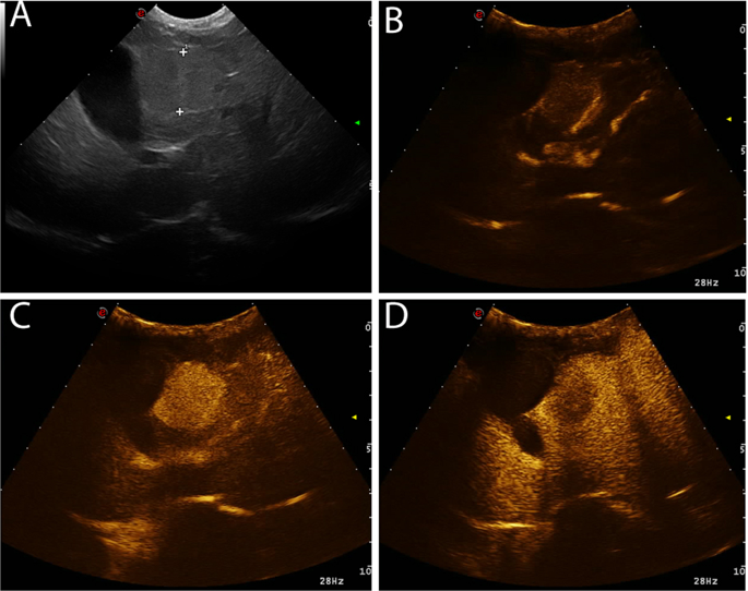
A total of 185 cases (150 retrospectively and 35 prospectively) of malignant liver masses were collected. In the retrospectively collected cases hyperenhancement during wash-in was the most common feature in HCCs but there was a high percentage of cases showing no enhancement or hypo/isoenhancement. ICCs displayed a large variety of contrast enhancement patterns and, although statically significant differences between ICCs and HCCs were evident, no clear distinction between these two pathologies was possible based only on their CEUS appearance. Sarcomas displayed all the possible degrees of wash-in enhancement with non-enhancing being the most common appearance. Metastases displayed all the possible contrast-enhancement patterns, with the most common being hyperenhancement in the wash-in phase followed by hypoenhancement in the wash-out phase. A decision tree was developed based on the features of the retrospectively selected cases. Based on the developed decision tree 27/35 prospectively collected cases were correctly classified. Even if some significant differences among groups were evident, all the histotypes displayed all the possible patterns of contrast enhancement, and, therefore, the differentiation of liver masses in dogs based only on their CEUS features is not feasible and, therefore, cytology or histopathology is required.
中文翻译:
犬恶性局灶性肝脏肿块的超声造影特征。
总共收集了185例(回顾性150例,预期35例)的恶性肝脏肿块。在回顾性收集的病例中,在清洗过程中过度增强是肝癌的最常见特征,但有很大比例的病例显示没有增强或低/等增强。ICC显示了各种各样的对比度增强模式,尽管ICC和HCC在静态上存在明显的显着差异,但仅根据其CEUS外观,无法将这两种病理学区分开。肉瘤显示出所有可能的增强浸润程度,其中最常见的表现是不增强。转移灶显示了所有可能的对比度增强模式,最常见的是在洗入阶段出现过度增强,然后在洗出阶段出现过度增强。基于回顾性选择案例的特征,开发了决策树。基于制定的决策树,正确分类了27/35个预期收集的病例。即使各组之间存在明显的显着差异,但所有组织型均显示出所有可能的对比度增强模式,因此仅依靠CEUS特征区分犬肝肿块是不可行的,因此,细胞学或组织病理学是可行的。需要。


 京公网安备 11010802027423号