Journal of Neuroradiology ( IF 3.5 ) Pub Date : 2020-03-10 , DOI: 10.1016/j.neurad.2020.03.001 Baojian Wang 1 , Tingqin Yan 2 , Jian Zhou 3 , Yuanzhong Xie 3 , Jianfeng Qiu 4 , Yi Wang 5 , Weizhao Lu 4
|
Background
High-tension glaucoma (HTG) is associated with functional changes in the brain, and elevated intraocular pressure (IOP) is one of the major causes.
Purpose
To evaluate the effects of high IOP on the brain in patients with HTG by using resting-state functional magnetic resonance imaging (rs-fMRI).
Materials and methods
Thirty-six patients with HTG and 20 age- and gender-matched healthy controls (HCs) were recruited and underwent IOP examination and rs-fMRI scan. Voxel-wise functional connectivity (FC) values were obtained between the Brodmann Area (BA) 17 (primary visual cortex) and the rest of the brain, two-sample t test was performed between HTG group and HCs. Correlation analysis was performed between FC and clinical information.
Results
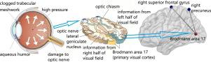
Compared with HCs, HTG patients demonstrated decreased FC between BA 17 and the right precuneus gyrus, decreased FC between BA 17 and the right superior frontal gyrus (SFG) (GRF corrected at voxel level P < 0.001 and cluster level P < 0.05, two-tailed). FC between BA 17 and the right SFG showed significantly negative correlation with right eyes’ IOP and mean IOP.
Conclusion
HTG patients had abnormal FC changes between the visual cortex and multiple functional brain regions related to visual sense, memory consolidation and cognitive processing, which provided image support for the pathophysiology research of HTG, and revealed new targets for the accurate treatment of HTG.
中文翻译:
高血压青光眼患者 fMRI 衍生功能连接的改变
背景
高血压青光眼 (HTG) 与大脑的功能变化有关,眼内压 (IOP) 升高是主要原因之一。
目的
使用静息态功能磁共振成像 (rs-fMRI) 评估高眼压对 HTG 患者大脑的影响。
材料和方法
招募了 36 名 HTG 患者和 20 名年龄和性别匹配的健康对照 (HC),并接受了眼压检查和 rs-fMRI 扫描。在布罗德曼区 (BA) 17 (初级视觉皮层) 和大脑的其余部分之间获得了体素功能连接 (FC) 值,在 HTG 组和 HCs 之间进行了两样本 t 检验。在FC和临床信息之间进行相关分析。
结果
与 HC 相比,HTG 患者表现出 BA 17 和右侧楔前叶之间的 FC 降低,BA 17 和右侧额上回 (SFG) 之间的 FC 降低(GRF 在体素水平P < 0.001 和簇水平P < 0.05校正,两尾)。BA 17 和右侧 SFG 之间的 FC 与右眼的 IOP 和平均 IOP 呈显着负相关。
结论
HTG患者视皮层与视觉、记忆巩固和认知处理相关的多个功能脑区之间存在异常的FC变化,为HTG的病理生理研究提供了图像支持,为HTG的精准治疗揭示了新的靶点。


 京公网安备 11010802027423号