当前位置:
X-MOL 学术
›
Acta Biomater.
›
论文详情
Our official English website, www.x-mol.net, welcomes your feedback! (Note: you will need to create a separate account there.)
Mesenchymal stem cell-laden, personalized 3D scaffolds with controlled structure and fiber alignment promote diabetic wound healing.
Acta Biomaterialia ( IF 9.7 ) Pub Date : 2020-04-05 , DOI: 10.1016/j.actbio.2020.03.035 Shixuan Chen 1 , Hongjun Wang 1 , Yajuan Su 1 , Johnson V John 1 , Alec McCarthy 1 , Shannon L Wong 2 , Jingwei Xie 1
Acta Biomaterialia ( IF 9.7 ) Pub Date : 2020-04-05 , DOI: 10.1016/j.actbio.2020.03.035 Shixuan Chen 1 , Hongjun Wang 1 , Yajuan Su 1 , Johnson V John 1 , Alec McCarthy 1 , Shannon L Wong 2 , Jingwei Xie 1
Affiliation
|
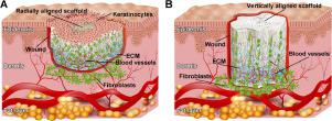
The management of diabetic wounds remains a major therapeutic challenge in clinics. Herein, we report a personalized treatment using 3D scaffolds consisting of radially or vertically aligned nanofibers in combination with bone marrow mesenchymal stem cells (BMSCs). The 3D scaffolds have customizable sizes, depths, and shapes, enabling them to fit a variety of type 2 diabetic wounds. In addition, the 3D scaffolds are shape-recoverable in atmosphere and water following compression. The BMSCs-laden 3D scaffolds are capable of enhancing the formation of granulation tissue, promoting angiogenesis, and facilitating collagen deposition. Further, such scaffolds inhibit the formation of M1-type macrophages and the expression of pro-inflammatory cytokines IL-6 and TNF-α and promote the formation of M2-type macrophages and the expression of anti-inflammatory cytokines IL-4 and IL-10. Taken together, BMSCs-laden, 3D nanofiber scaffolds with controlled structure and alignment hold great promise for the treatment of diabetic wounds. STATEMENT OF SIGNIFICANCE: In this study, we developed 3D radially and vertically aligned nanofiber scaffolds to transplant bone marrow mesenchymal stem cells (BMSCs). We personalized 3D scaffolds that could completely match the size, depth, and shape of diabetic wounds. Moreover, both the radially and vertically aligned nanofiber scaffolds could completely recover their shape and maintain structural integrity after repeated loads with compressive stresses. Furthermore, the BMSCs-laden 3D scaffolds are able to promote granulation tissue formation, angiogenesis, and collagen deposition, and switch the immune responses to the pro-regenerative direction. These 3D scaffolds consisting of radially or vertically aligned nanofibers in combination with BMSCs offer a robust, customizable platform potentially for a significant improvement of managing diabetic wounds.
中文翻译:

间充质干细胞,个性化的3D支架具有受控的结构和纤维排列,可促进糖尿病伤口的愈合。
糖尿病伤口的管理仍然是临床上的主要治疗挑战。本文中,我们报告了使用3D支架进行个性化治疗的方法,该支架由径向或垂直排列的纳米纤维与骨髓间充质干细胞(BMSC)组成。3D支架具有可自定义的大小,深度和形状,使它们能够适合各种2型糖尿病伤口。此外,3D支架在压缩后可在大气和水中恢复形状。装有BMSCs的3D支架能够增强肉芽组织的形成,促进血管生成并促进胶原蛋白的沉积。进一步,这样的支架抑制M1型巨噬细胞的形成和促炎细胞因子IL-6和TNF-α的表达,并促进M2型巨噬细胞的形成以及抗炎细胞因子IL-4和IL-10的表达。总之,具有受控结构和排列的载满BMSCs的3D纳米纤维支架有望为糖尿病伤口的治疗提供前景。重大意义声明:在这项研究中,我们开发了3D径向和垂直对齐的纳米纤维支架,以移植骨髓间充质干细胞(BMSC)。我们对3D支架进行了个性化设置,可以完全匹配糖尿病伤口的大小,深度和形状。此外,在反复施加压缩应力后,径向和垂直排列的纳米纤维支架都可以完全恢复其形状并保持结构完整性。此外,载有BMSCs的3D支架能够促进肉芽组织形成,血管生成和胶原蛋白沉积,并将免疫反应切换至促再生方向。这些由3D径向或垂直排列的纳米纤维与BMSC组成的3D支架提供了强大的,可定制的平台,可能显着改善糖尿病伤口的管理。
更新日期:2020-04-05
中文翻译:
间充质干细胞,个性化的3D支架具有受控的结构和纤维排列,可促进糖尿病伤口的愈合。
糖尿病伤口的管理仍然是临床上的主要治疗挑战。本文中,我们报告了使用3D支架进行个性化治疗的方法,该支架由径向或垂直排列的纳米纤维与骨髓间充质干细胞(BMSC)组成。3D支架具有可自定义的大小,深度和形状,使它们能够适合各种2型糖尿病伤口。此外,3D支架在压缩后可在大气和水中恢复形状。装有BMSCs的3D支架能够增强肉芽组织的形成,促进血管生成并促进胶原蛋白的沉积。进一步,这样的支架抑制M1型巨噬细胞的形成和促炎细胞因子IL-6和TNF-α的表达,并促进M2型巨噬细胞的形成以及抗炎细胞因子IL-4和IL-10的表达。总之,具有受控结构和排列的载满BMSCs的3D纳米纤维支架有望为糖尿病伤口的治疗提供前景。重大意义声明:在这项研究中,我们开发了3D径向和垂直对齐的纳米纤维支架,以移植骨髓间充质干细胞(BMSC)。我们对3D支架进行了个性化设置,可以完全匹配糖尿病伤口的大小,深度和形状。此外,在反复施加压缩应力后,径向和垂直排列的纳米纤维支架都可以完全恢复其形状并保持结构完整性。此外,载有BMSCs的3D支架能够促进肉芽组织形成,血管生成和胶原蛋白沉积,并将免疫反应切换至促再生方向。这些由3D径向或垂直排列的纳米纤维与BMSC组成的3D支架提供了强大的,可定制的平台,可能显着改善糖尿病伤口的管理。


 京公网安备 11010802027423号