Journal of the Mechanical Behavior of Biomedical Materials ( IF 3.9 ) Pub Date : 2020-04-03 , DOI: 10.1016/j.jmbbm.2020.103761 Daniela Scherrer 1 , Urs Bragger 1 , Marco Ferrari 2 , André Mocker 3 , Tim Joda 4
|
Aim
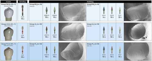
The objective of this In-vitro investigation was to analyze and compare the surface after polishing of 63 all-ceramic restorations fabricated out of monolithic zirconium dioxide (ZIR), lithium disilicate (LS) or feldspathic ceramic (FC) under standardized laboratory conditions with different protocols. The primary outcome was defined as the roughness (Ra/Sa) of different ceramic surfaces after distinctive polishing procedures.
Material and methods
The study set-up consisted of three main groups: ZIR, LS, FC (20 crowns each), every group divided into two sub-groups (10 crowns each) depending on the polishing method. The untouched glazed surface of one crown per material served as a control. Every crown displayed a defined supra-contact at the palatal cusp which was removed with a fine grain (38–45 μm) diamond bur. Surface polishing was carried out with either a two-step system (one kit for zirconium dioxide (ZIR2), another kit for lithium disilicate (LS2) and feldspatic ceramic (FC2)), or a three-step system (ZIR3, LS3, FC3) under standardized conditions. Roughness parameters (Ra and Sa) were measured by means of confocal profilometry. Specimens were also visually inspected with scanning electron microscopy (SEM). Statistical analyses were performed using SPSS Statistics software.
Results
Visual examination of the specimens using SEM showed several inhomogeneities on the glazed surface of the control samples, i.e. pores and particles. On every test sample, the grinding curves of the diamond bur were still recognizable. Polishing revealed similar median Ra 0.491 μm (ZIR2) and 0.434 μm (LS2) after two-step polishing (p = 0.754), and 0.311 μm (ZIR3) and 0.208 μm (LS3) after three-step polishing (p = 0.917). Surface roughness in group FC measured 0.889 μm (FC2) after the two-step polishing process and 0.903 μm (FC3) following three-step surface refinement. No significant difference was detectable between surface roughness of glazed controls compared to either polished surfaces with two-step or three-step treatment within one material. ZIR and LS presented significantly lower median roughness Ra after two-step and three-step procedures than test samples of FC, measured subsequent to either of the polishing methods (p = 0.016, p = 0.009).
Conclusion
The surface roughness of ZIR, LS and FC crowns after the use of chairside polishing kits was comparable with the roughness measured before occlusal adjustment. A two-step procedure showed as good results as a three-step process. A smoother surface was obtained for ZIR and LS compared to FC with both polishing protocols.
中文翻译:
CAD / CAM陶瓷修复体的体外抛光:使用SEM和共聚焦轮廓仪进行评估。
目标
这项体外研究的目的是分析和比较在标准化实验室条件下用不同的二氧化锆(ZIR),二硅酸锂(LS)或长石陶瓷(FC)制成的63种全陶瓷修复体的抛光后表面。协议。主要结果定义为经过独特抛光程序后不同陶瓷表面的粗糙度(R a / S a)。
材料与方法
研究设置包括三个主要组:ZIR,LS,FC(每个20冠),每个组根据抛光方法分为两个子组(每个10冠)。每种材料的一个表冠未打磨的釉面作为对照。每个牙冠在the尖处都显示出明确的超接触,并用细颗粒(38–45μm)的钻石钻将其去除。表面抛光可以通过两步系统(一个用于二氧化锆的套件(ZIR 2),另一个用于二硅酸锂(LS 2)和弹性陶瓷(FC 2)的套件)或一个三步系统(ZIR 3)进行。,LS 3,FC 3)。粗糙度参数(R a和S a)通过共聚焦轮廓仪测量。还用扫描电子显微镜(SEM)目测检查样品。使用SPSS Statistics软件进行统计分析。
结果
使用SEM对样品进行的目视检查显示,对照样品的釉面存在多个不均匀性,即孔和颗粒。在每个测试样品上,金刚石车针的磨削曲线仍然可以识别。抛光显示两步抛光(p = 0.754)后的中值R a分别为0.491μm(ZIR 2)和0.434μm (LS 2),三步抛光后的中位数R a为0.311μm (ZIR 3)和0.208μm (LS 3)(p = 0.754)p = 0.917)。经过两步抛光后,FC组的表面粗糙度为0.889μm (FC 2),而表面粗糙度为0.903μm(FC 3)进行三步表面精修。与在一种材料中经过两步或三步处理的抛光表面相比,上釉对照的表面粗糙度之间没有可检测到的显着差异。ZIR和LS在经过两步和三步程序后呈现出比FC的测试样品低得多的中值粗糙度R a,而这两种抛光方法中的任一种都经过测试(p = 0.016,p = 0.009)。
结论
使用椅背抛光套件后,ZIR,LS和FC牙冠的表面粗糙度与咬合调整前测得的粗糙度相当。两步过程显示出与三步过程一样好的结果。与两种抛光方案的FC相比,ZIR和LS均获得了更光滑的表面。


 京公网安备 11010802027423号