当前位置:
X-MOL 学术
›
Nanoscale Adv.
›
论文详情
Our official English website, www.x-mol.net, welcomes your feedback! (Note: you will need to create a separate account there.)
Cellular regeneration and proliferation on polymeric 3D inverse-space substrates and the effect of doxorubicin
Nanoscale Advances ( IF 4.7 ) Pub Date : 2020-04-01 , DOI: 10.1039/d0na00075b Chandrashekhar D Bobade 1 , Semonti Nandi 1 , Narendra R Kale 1 , Shashwat S Banerjee 2 , Yuvraj N Patil 2 , Jayant J Khandare 3
Nanoscale Advances ( IF 4.7 ) Pub Date : 2020-04-01 , DOI: 10.1039/d0na00075b Chandrashekhar D Bobade 1 , Semonti Nandi 1 , Narendra R Kale 1 , Shashwat S Banerjee 2 , Yuvraj N Patil 2 , Jayant J Khandare 3
Affiliation
|
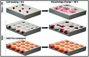
Spatial arrangement for cells and the opportunity thereof have implications in cell regeneration and cell proliferation. 3D inverse space (3DIS) substrates with micron-sized pores are fabricated under controlled environmental conditions from polymers such as poly(lactic-co-glycolic) acid (PLGA), poly(lactic acid) (PLA) and poly(styrene) (PS). The characterization of 3DIS substrates by optical microscopy, scanning probe microscopy (SPM), etc. shows pores within 1–18 μm diameter and prominent surface roughness extending up to 3.9 nm in height over its base. Conversely, to compare two-dimensional (2D) versus 3DIS substrates, the crucial variables of cell height, cell spreading area and cell volume are compared using lung adenocarcinoma (A549) cells. The results indicate an average cell thickness of ∼6 μm on a glass substrate whereas cells on PLGA 3DIS were ∼12 μm in height, occasionally reaching 20 μm, with a 40% decreased cell spreading area. A549 cells cultured on polymer 3DIS substrates show a cell regeneration growth pattern, dependent on the available spatial volume. Furthermore, PLGA 3DIS cell culture systems with and without graded doxorubicin (DOX) pre-treatment result in potent cell inhibition and cell proliferation, respectively. Additionally, standard DOX administration to A549 cells in the PLGA 3DIS system revealed altered drug sensitivity. 3DIS demonstrates utility in facilitating cellular regeneration and mimicking cell proliferation in defined spatial arrangements.
中文翻译:

聚合物3D逆空间底物的细胞再生和增殖及多柔比星的作用
细胞的空间排列及其机会对细胞再生和细胞增殖有影响。具有微米级孔隙的 3D 逆空间 (3DIS) 基板是在受控环境条件下由聚(乳酸-共-乙醇)酸(PLGA)、聚(乳酸)(PLA)和聚(苯乙烯)(PS)等聚合物制造的)。通过光学显微镜、扫描探针显微镜 (SPM)等对 3DIS 基板进行表征,显示直径在 1-18 μm 范围内的孔和突出的表面粗糙度,在其底部延伸高达 3.9 nm。相反,比较二维 (2D)与使用肺腺癌细胞 (A549) 比较 3DIS 底物、细胞高度、细胞扩散面积和细胞体积的关键变量。结果表明,玻璃基板上的平均细胞厚度约为 6 μm,而 PLGA 3DIS 上的细胞高度约为 12 μm,偶尔达到 20 μm,细胞扩散面积减少了 40%。在聚合物 3DIS 基质上培养的 A549 细胞显示出细胞再生生长模式,这取决于可用的空间体积。此外,具有和不具有分级多柔比星 (DOX) 预处理的 PLGA 3DIS 细胞培养系统分别导致有效的细胞抑制和细胞增殖。此外,在 PLGA 3DIS 系统中对 A549 细胞的标准 DOX 给药揭示了药物敏感性的改变。
更新日期:2020-04-01
中文翻译:
聚合物3D逆空间底物的细胞再生和增殖及多柔比星的作用
细胞的空间排列及其机会对细胞再生和细胞增殖有影响。具有微米级孔隙的 3D 逆空间 (3DIS) 基板是在受控环境条件下由聚(乳酸-共-乙醇)酸(PLGA)、聚(乳酸)(PLA)和聚(苯乙烯)(PS)等聚合物制造的)。通过光学显微镜、扫描探针显微镜 (SPM)等对 3DIS 基板进行表征,显示直径在 1-18 μm 范围内的孔和突出的表面粗糙度,在其底部延伸高达 3.9 nm。相反,比较二维 (2D)与使用肺腺癌细胞 (A549) 比较 3DIS 底物、细胞高度、细胞扩散面积和细胞体积的关键变量。结果表明,玻璃基板上的平均细胞厚度约为 6 μm,而 PLGA 3DIS 上的细胞高度约为 12 μm,偶尔达到 20 μm,细胞扩散面积减少了 40%。在聚合物 3DIS 基质上培养的 A549 细胞显示出细胞再生生长模式,这取决于可用的空间体积。此外,具有和不具有分级多柔比星 (DOX) 预处理的 PLGA 3DIS 细胞培养系统分别导致有效的细胞抑制和细胞增殖。此外,在 PLGA 3DIS 系统中对 A549 细胞的标准 DOX 给药揭示了药物敏感性的改变。


 京公网安备 11010802027423号