PLOS ONE ( IF 3.7 ) Pub Date : 2020-03-31 , DOI: 10.1371/journal.pone.0230553 Johannes Thüring 1 , Christiane Katharina Kuhl 1 , Alexandra Barabasch 1 , Lea Hitpass 1 , Maike Bode 1 , Nina Bünting 1 , Philipp Bruners 1 , Nils Andreas Krämer 1
|
Objective
The purpose of this study was to investigate signal changes in T2-weighted magnetic resonance imaging of liver metastases under treatment with and without bevacizumab-containing chemotherapy and to compare these signal changes to tumor contrast enhancement.
Materials and methods
Retrospective analysis of 44 patients, aged 36–84 years, who underwent liver magnetic resonance imaging including T2-weighted and dynamic contrast enhancement sequences. Patients received bevacizumab-containing (n = 22) or conventional cytotoxic chemotherapy (n = 22). Magnetic resonance imaging was obtained at baseline and at three follow-ups (on average 3, 6 and 9 months after initial treatment). Three independent readers rated the T2 signal intensity and the relative contrast enhancement of the metastases on a 5-point scale.
Results
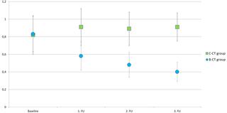
T2 signal intensity of metastases treated with bevacizumab showed a significant (p<0.001) decrease in T2 signal intensity after initial treatment and exhibit compared to conventionally treated metastases significantly (p<0.001 for each follow-up) hypointense (bevacizumab: 0.70 ± 0.83 before vs. -1.55 ± 0.61, -1.91 ± 0.62, and -1.97 ± 0.52; cytotoxic: 0.73 ± 0.79 before vs. -0.69 ± 0.81, -0.71 ± 0.68, and -0.75 ± 0.65 after 3, 6, and 9 months, respectively). T2 signal intensity was strongly correlated with tumor contrast enhancement (r = 0.71; p<0.001). Intra-observer agreement for T2-signal intensity was substantial (κ = 0.75). The agreement for tumoral contrast enhancement between the readers was considerably lower (κ = 0.39).
Conclusion
Liver metastases exhibit considerably hypointense in T2-weighted imaging after treatment with bevacizumab, in contrast to conventionally treated liver metastases. Therefore, T2-weighted imaging seems to reflect the effect of bevacizumab.
中文翻译:
贝伐单抗-一种实用的成像生物标记物在肝转移的T2加权MRI中的信号变化?
目的
这项研究的目的是调查在接受和不接受含贝伐单抗的化学疗法治疗下肝转移的T2加权磁共振成像中的信号变化,并将这些信号变化与肿瘤造影剂增强进行比较。
材料和方法
回顾性分析了44例年龄在36-84岁的患者,他们接受了包括T2加权和动态对比增强序列在内的肝脏磁共振成像。患者接受含贝伐单抗的治疗(n = 22)或常规细胞毒性化疗(n = 22)。在基线和三个随访(初次治疗后平均3、6和9个月)获得磁共振成像。三个独立的阅读器以5分制对T2信号强度和转移灶的相对对比度增强进行了评分。
结果
与贝伐单抗治疗转移的T2信号强度显示出显著(P <0.001)减少T2信号强度初始治疗并表现出后相比显著常规处理转移(p <0.001对于每个后续)低信号(贝伐单抗:0.70±0.83之前相比-1.55±0.61,-1.91±0.62和-1.97±0.52;细胞毒性:3个月,6个月和9个月之前为0.73±0.79,而-0.69±0.81,-0.71±0.68和-0.75±0.65分别)。T2信号强度与肿瘤造影剂增强密切相关(r = 0.71;p <0.001)。T2信号强度的观察者内部一致性很高(κ = 0.75)。读者之间的肿瘤对比度增强的一致性要低得多(κ = 0.39)。
结论
与常规治疗的肝转移瘤相比,贝伐单抗治疗后的肝转移瘤在T2加权成像中表现出相当低的凝结性。因此,T2加权成像似乎反映了贝伐单抗的作用。


 京公网安备 11010802027423号