当前位置:
X-MOL 学术
›
PLOS Pathog.
›
论文详情
Our official English website, www.x-mol.net, welcomes your feedback! (Note: you will need to create a separate account there.)
An improved animal model for herpesvirus encephalitis in humans.
PLoS Pathogens ( IF 6.7 ) Pub Date : 2020-03-30 , DOI: 10.1371/journal.ppat.1008445 Julia Sehl 1, 2 , Julia E Hölper 1 , Barbara G Klupp 1 , Christina Baumbach 3 , Jens P Teifke 2 , Thomas C Mettenleiter 1
PLoS Pathogens ( IF 6.7 ) Pub Date : 2020-03-30 , DOI: 10.1371/journal.ppat.1008445 Julia Sehl 1, 2 , Julia E Hölper 1 , Barbara G Klupp 1 , Christina Baumbach 3 , Jens P Teifke 2 , Thomas C Mettenleiter 1
Affiliation
|
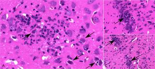
Herpesviral encephalitis caused by Herpes Simplex Virus 1 (HSV-1) is one of the most devastating diseases in humans. Patients present with fever, mental status changes or seizures and when untreated, sequelae can be fatal. Herpes Simplex Encephalitis (HSE) is characterized by mainly unilateral necrotizing inflammation effacing the frontal and mesiotemporal lobes with rare involvement of the brainstem. HSV-1 is hypothesized to invade the CNS via the trigeminal or olfactory nerve, but viral tropism and the exact route of infection remain unclear. Several mouse models for HSE have been developed, but they mimic natural infection only inadequately. The porcine alphaherpesvirus Pseudorabies virus (PrV) is closely related to HSV-1 and Varicella Zoster Virus (VZV). While pigs can control productive infection, it is lethal in other susceptible animals associated with severe pruritus leading to automutilation. Here, we describe the first mutant PrV establishing productive infection in mice that the animals are able to control. After intranasal inoculation with a PrV mutant lacking tegument protein pUL21 and pUS3 kinase activity (PrV-ΔUL21/US3Δkin), nearly all mice survived despite extensive infection of the central nervous system. Neuroinvasion mainly occurred along the trigeminal pathway. Whereas trigeminal first and second order neurons and autonomic ganglia were positive early after intranasal infection, PrV-specific antigen was mainly detectable in the frontal, mesiotemporal and parietal lobes at later times, accompanied by a long lasting lymphohistiocytic meningoencephalitis. Despite this extensive infection, mice showed only mild to moderate clinical signs, developed alopecic skin lesions, or remained asymptomatic. Interestingly, most mice exhibited abnormalities in behavior and activity levels including slow movements, akinesia and stargazing. In summary, clinical signs, distribution of viral antigen and inflammatory pattern show striking analogies to human encephalitis caused by HSV-1 or VZV not observed in other animal models of disease.
中文翻译:

改进的人类疱疹病毒性脑炎动物模型。
由单纯疱疹病毒1(HSV-1)引起的疱疹病毒性脑炎是人类中最具破坏性的疾病之一。出现发烧,精神状态改变或癫痫发作的患者,如果不及时治疗,后遗症可能是致命的。单纯疱疹性脑炎(HSE)的特征是主要出现在额叶和近颞叶的单侧坏死性炎症,很少累及脑干。假设HSV-1通过三叉神经或嗅神经侵入CNS,但尚不清楚病毒的嗜性和确切的感染途径。已经开发了几种HSE小鼠模型,但是它们仅不足以模仿自然感染。猪α疱疹病毒伪狂犬病病毒(PrV)与HSV-1和水痘带状疱疹病毒(VZV)密切相关。尽管猪可以控制生产性感染,它在与严重瘙痒相关的其他易感动物中具有致死作用,导致自残。在这里,我们描述了第一个突变的PrV在动物能够控制的小鼠中建立生产性感染。鼻内接种缺乏皮层蛋白pUL21和pUS3激酶活性(PrV-ΔUL21/US3Δkin)的PrV突变体后,尽管中枢神经系统受到了广泛感染,几乎所有小鼠都存活了下来。神经入侵主要发生在三叉神经途径上。鼻内感染后三叉神经的一,二阶神经元和自主神经节呈阳性,而PrV特异性抗原则主要在额叶,中颞叶和顶叶中检出,并伴有长时间的淋巴组织细胞性脑膜脑炎。尽管受到广泛感染,小鼠仅表现出轻度至中度的临床体征,发展成秃头症的皮肤病变或保持无症状。有趣的是,大多数小鼠表现出行为和活动水平异常,包括动作缓慢,运动障碍和注视。总之,临床体征,病毒抗原的分布和炎性模式显示出与在其他疾病模型中未观察到的由HSV-1或VZV引起的人脑炎的惊人相似之处。
更新日期:2020-03-30
中文翻译:
改进的人类疱疹病毒性脑炎动物模型。
由单纯疱疹病毒1(HSV-1)引起的疱疹病毒性脑炎是人类中最具破坏性的疾病之一。出现发烧,精神状态改变或癫痫发作的患者,如果不及时治疗,后遗症可能是致命的。单纯疱疹性脑炎(HSE)的特征是主要出现在额叶和近颞叶的单侧坏死性炎症,很少累及脑干。假设HSV-1通过三叉神经或嗅神经侵入CNS,但尚不清楚病毒的嗜性和确切的感染途径。已经开发了几种HSE小鼠模型,但是它们仅不足以模仿自然感染。猪α疱疹病毒伪狂犬病病毒(PrV)与HSV-1和水痘带状疱疹病毒(VZV)密切相关。尽管猪可以控制生产性感染,它在与严重瘙痒相关的其他易感动物中具有致死作用,导致自残。在这里,我们描述了第一个突变的PrV在动物能够控制的小鼠中建立生产性感染。鼻内接种缺乏皮层蛋白pUL21和pUS3激酶活性(PrV-ΔUL21/US3Δkin)的PrV突变体后,尽管中枢神经系统受到了广泛感染,几乎所有小鼠都存活了下来。神经入侵主要发生在三叉神经途径上。鼻内感染后三叉神经的一,二阶神经元和自主神经节呈阳性,而PrV特异性抗原则主要在额叶,中颞叶和顶叶中检出,并伴有长时间的淋巴组织细胞性脑膜脑炎。尽管受到广泛感染,小鼠仅表现出轻度至中度的临床体征,发展成秃头症的皮肤病变或保持无症状。有趣的是,大多数小鼠表现出行为和活动水平异常,包括动作缓慢,运动障碍和注视。总之,临床体征,病毒抗原的分布和炎性模式显示出与在其他疾病模型中未观察到的由HSV-1或VZV引起的人脑炎的惊人相似之处。


 京公网安备 11010802027423号