当前位置:
X-MOL 学术
›
Nat. Commun.
›
论文详情
Our official English website, www.x-mol.net, welcomes your feedback! (Note: you will need to create a separate account there.)
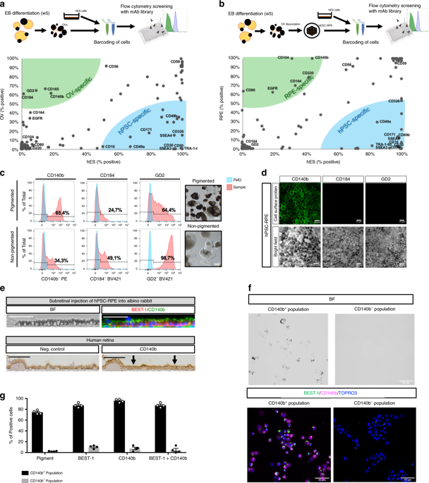
Identification of cell surface markers and establishment of monolayer differentiation to retinal pigment epithelial cells.
Nature Communications ( IF 16.6 ) Pub Date : 2020-03-30 , DOI: 10.1038/s41467-020-15326-5 Alvaro Plaza Reyes 1, 2, 3 , Sandra Petrus-Reurer 1, 2, 3, 4 , Sara Padrell Sánchez 1, 2, 3 , Pankaj Kumar 1, 2, 3 , Iyadh Douagi 5 , Hammurabi Bartuma 4 , Monica Aronsson 4 , Sofie Westman 4 , Emma Lardner 4 , Helder André 4 , Anna Falk 6 , Emeline F Nandrot 7 , Anders Kvanta 4 , Fredrik Lanner 1, 2, 3
Nature Communications ( IF 16.6 ) Pub Date : 2020-03-30 , DOI: 10.1038/s41467-020-15326-5 Alvaro Plaza Reyes 1, 2, 3 , Sandra Petrus-Reurer 1, 2, 3, 4 , Sara Padrell Sánchez 1, 2, 3 , Pankaj Kumar 1, 2, 3 , Iyadh Douagi 5 , Hammurabi Bartuma 4 , Monica Aronsson 4 , Sofie Westman 4 , Emma Lardner 4 , Helder André 4 , Anna Falk 6 , Emeline F Nandrot 7 , Anders Kvanta 4 , Fredrik Lanner 1, 2, 3
Affiliation
|
In vitro differentiation of human pluripotent stem cells into functional retinal pigment epithelial (RPE) cells provides a potentially unlimited source for cell based reparative therapy of age-related macular degeneration. Although the inherent pigmentation of the RPE cells have been useful to grossly evaluate differentiation efficiency and allowed manual isolation of pigmented structures, accurate quantification and automated isolation has been challenging. To address this issue, here we perform a comprehensive antibody screening and identify cell surface markers for RPE cells. We show that these markers can be used to isolate RPE cells during in vitro differentiation and to track, quantify and improve differentiation efficiency. Finally, these surface markers aided to develop a robust, direct and scalable monolayer differentiation protocol on human recombinant laminin-111 and -521 without the need for manual isolation.
中文翻译:

鉴定细胞表面标志物并建立视网膜色素上皮细胞的单层分化。
人多能干细胞在体外分化为功能性视网膜色素上皮(RPE)细胞为年龄相关性黄斑变性的细胞修复治疗提供了潜在的无限来源。尽管RPE细胞固有的色素沉着可用于全面评估分化效率并允许手动隔离色素结构,但准确的定量和自动隔离仍具有挑战性。为了解决这个问题,我们在这里进行全面的抗体筛选并鉴定RPE细胞的细胞表面标记。我们表明,这些标记物可用于在体外分化过程中分离RPE细胞,以及跟踪,量化和提高分化效率。最后,这些表面标记有助于形成坚固,
更新日期:2020-04-24
中文翻译:
鉴定细胞表面标志物并建立视网膜色素上皮细胞的单层分化。
人多能干细胞在体外分化为功能性视网膜色素上皮(RPE)细胞为年龄相关性黄斑变性的细胞修复治疗提供了潜在的无限来源。尽管RPE细胞固有的色素沉着可用于全面评估分化效率并允许手动隔离色素结构,但准确的定量和自动隔离仍具有挑战性。为了解决这个问题,我们在这里进行全面的抗体筛选并鉴定RPE细胞的细胞表面标记。我们表明,这些标记物可用于在体外分化过程中分离RPE细胞,以及跟踪,量化和提高分化效率。最后,这些表面标记有助于形成坚固,


 京公网安备 11010802027423号