当前位置:
X-MOL 学术
›
Toxicol. Appl. Pharmacol.
›
论文详情
Our official English website, www.x-mol.net, welcomes your feedback! (Note: you will need to create a separate account there.)
Aldehyde dehydrogenase-2 activation decreases acetaminophen hepatotoxicity by prevention of mitochondrial depolarization.
Toxicology and Applied Pharmacology ( IF 3.8 ) Pub Date : 2020-03-30 , DOI: 10.1016/j.taap.2020.114982 Hereward J Wimborne 1 , Jiangting Hu 1 , Kenji Takemoto 1 , Nga T Nguyen 2 , Hartmut Jaeschke 2 , John J Lemasters 3 , Zhi Zhong 1
Toxicology and Applied Pharmacology ( IF 3.8 ) Pub Date : 2020-03-30 , DOI: 10.1016/j.taap.2020.114982 Hereward J Wimborne 1 , Jiangting Hu 1 , Kenji Takemoto 1 , Nga T Nguyen 2 , Hartmut Jaeschke 2 , John J Lemasters 3 , Zhi Zhong 1
Affiliation
|
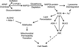
Oxidative stress contributes to acetaminophen (APAP) hepatotoxicity. Since lipid peroxidation produces reactive aldehydes, we investigated whether activation of mitochondrial aldehyde dehydrogenase-2 (ALDH2) with Alda-1 decreases liver injury after APAP. Male C57BL/6 mice fasted overnight received Alda-1 (20 mg/kg, i.p.) or vehicle 30 min before APAP (300 mg/kg, i.p.). Blood and livers were collected 2 or 24 h after APAP. Intravital multiphoton microscopy of rhodamine 123 (Rh123) and propidium iodide (PI) fluorescence was conducted 6 h after APAP administration to detect mitochondrial polarization status and cell death. 4-Hydroxynonenal protein adducts were present in 0.1% of tissue area without APAP treatment but increased to 7% 2 h after APAP treatment, which Alda-1 blunted to 1%. Serum alanine and aspartate aminotransferases increased to 7594 and 9768 U/L at 24 h respectively, which decreased ≥72% by Alda-1. Alda-1 also decreased centrilobular necrosis at 24 h after APAP from 47% of lobular areas to 21%. N-acetyl-p-benzoquinone imine protein adduct formation and c-Jun-N-terminal kinase phosphorylation increased after APAP as expected, but Alda-1 did not alter these changes. Without APAP, no mitochondrial depolarization was detected by intravital microscopy. At 6 h after APAP, 62% of tissue area showed depolarization, which decreased to 33.5% with Alda-1. Cell death as detected by PI labeling increased from 0 to 6.8 cells per 30× field 6 h after APAP, which decreased to 0.6 cells by Alda-1. In conclusion, aldehydes are important mediators of APAP hepatotoxicity. Accelerated aldehyde degradation by ALDH2 activation with Alda-1 decreases APAP hepatotoxicity by protection against mitochondrial dysfunction.
中文翻译:

醛脱氢酶2激活可通过防止线粒体去极化来降低对乙酰氨基酚的肝毒性。
氧化应激会导致对乙酰氨基酚(APAP)肝毒性。由于脂质过氧化会产生活性醛,因此我们研究了用Alda-1激活线粒体醛脱氢酶2(ALDH2)是否能减少APAP后的肝损伤。禁食过夜的雄性C57BL / 6小鼠在APAP前30分钟接受Alda-1(20 mg / kg,ip)或溶媒(300 mg / kg,ip)。APAP后2或24小时收集血液和肝脏。APAP给药后6小时进行了若丹明123(Rh123)和碘化丙啶(PI)荧光的活体多光子显微镜检查,以检测线粒体的极化状态和细胞死亡。未经APAP处理的组织中0.1%存在4-羟基壬醛蛋白加合物,但在APAP处理后2 h增加到7%,Alda-1减弱至1%。血清丙氨酸和天冬氨酸转氨酶在24 h分别升高至7594和9768 U / L,而Alda-1降低≥72%。Alda-1还可以在APAP后24小时将小叶坏死从47%的小叶区域减少到21%。APAP后,N-乙酰基-对-苯醌亚胺亚胺蛋白加合物的形成和c-Jun-N-端激酶的磷酸化增加,但Alda-1并未改变这些变化。没有APAP,通过活体内显微镜检查未检测到线粒体去极化。APAP后6小时,有62%的组织区域显示去极化,而Alda-1减少到33.5%。通过PI标记检测到的细胞死亡在APAP后6小时每30x场从0增加到6.8细胞,而通过Alda-1减少到0.6细胞。总之,醛是APAP肝毒性的重要介质。
更新日期:2020-03-31
中文翻译:
醛脱氢酶2激活可通过防止线粒体去极化来降低对乙酰氨基酚的肝毒性。
氧化应激会导致对乙酰氨基酚(APAP)肝毒性。由于脂质过氧化会产生活性醛,因此我们研究了用Alda-1激活线粒体醛脱氢酶2(ALDH2)是否能减少APAP后的肝损伤。禁食过夜的雄性C57BL / 6小鼠在APAP前30分钟接受Alda-1(20 mg / kg,ip)或溶媒(300 mg / kg,ip)。APAP后2或24小时收集血液和肝脏。APAP给药后6小时进行了若丹明123(Rh123)和碘化丙啶(PI)荧光的活体多光子显微镜检查,以检测线粒体的极化状态和细胞死亡。未经APAP处理的组织中0.1%存在4-羟基壬醛蛋白加合物,但在APAP处理后2 h增加到7%,Alda-1减弱至1%。血清丙氨酸和天冬氨酸转氨酶在24 h分别升高至7594和9768 U / L,而Alda-1降低≥72%。Alda-1还可以在APAP后24小时将小叶坏死从47%的小叶区域减少到21%。APAP后,N-乙酰基-对-苯醌亚胺亚胺蛋白加合物的形成和c-Jun-N-端激酶的磷酸化增加,但Alda-1并未改变这些变化。没有APAP,通过活体内显微镜检查未检测到线粒体去极化。APAP后6小时,有62%的组织区域显示去极化,而Alda-1减少到33.5%。通过PI标记检测到的细胞死亡在APAP后6小时每30x场从0增加到6.8细胞,而通过Alda-1减少到0.6细胞。总之,醛是APAP肝毒性的重要介质。


 京公网安备 11010802027423号