Laboratory Investigation ( IF 5 ) Pub Date : 2020-03-23 , DOI: 10.1038/s41374-020-0417-4 Jason Ptacek 1 , Darren Locke 2 , Rachel Finck 1 , Mary-Ellen Cvijic 2 , Zhuyin Li 2 , Jay G Tarolli 1 , Murat Aksoy 1 , Yari Sigal 1 , Yi Zhang 1 , Matt Newgren 1 , Jessica Finn 1
|
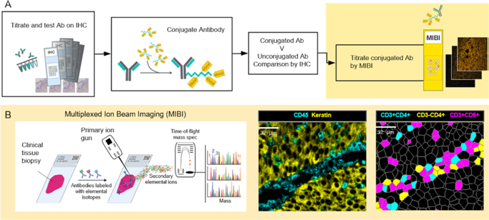
An ability to characterize the cellular composition and spatial organization of the tumor microenvironment (TME) using multiplexed IHC has been limited by the techniques available. Here we show the applicability of multiplexed ion beam imaging (MIBI) for cell phenotype identification and analysis of spatial relationships across numerous tumor types. Formalin-fixed paraffin-embedded (FFPE) samples from tumor biopsies were simultaneously stained with a panel of 15 antibodies, each labeled with a specific metal isotope. Multi-step processing produced images of the TME that were further segmented into single cells. Frequencies of different cell subsets and the distributions of nearest neighbor distances between them were calculated using this data. A total of 50 tumor specimens from 15 tumor types were characterized for their immune profile and spatial organization. Most samples showed infiltrating cytotoxic T cells and macrophages present amongst tumor cells. Spatial analysis of the TME in two ovarian serous carcinoma images highlighted differences in the degree of mixing between tumor and immune cells across samples. Identification of admixed PD-L1+ macrophages and PD-1+ T cells in an urothelial carcinoma sample allowed for the detailed observations of immune cell subset spatial arrangement. These results illustrate the high-parameter capability of MIBI at a sensitivity and resolution uniquely suited to understanding the complex tumor immune landscape including the spatial relationships of immune and tumor cells and expression of immunoregulatory proteins.
中文翻译:
多路离子束成像 (MIBI),用于表征不同肿瘤类型的肿瘤微环境。
使用多重 IHC 表征肿瘤微环境 (TME) 的细胞组成和空间组织的能力受到可用技术的限制。在这里,我们展示了多重离子束成像 (MIBI) 在细胞表型识别和多种肿瘤类型空间关系分析中的适用性。来自肿瘤活检的福尔马林固定石蜡包埋 (FFPE) 样本同时用一组 15 种抗体染色,每种抗体都标记有特定的金属同位素。多步处理产生了 TME 的图像,这些图像被进一步分割成单个细胞。使用该数据计算不同细胞子集的频率和它们之间最近邻距离的分布。对来自 15 种肿瘤类型的总共 50 个肿瘤标本进行了免疫特征和空间组织的表征。大多数样本显示肿瘤细胞中存在浸润性细胞毒性 T 细胞和巨噬细胞。两张卵巢浆液性癌图像中 TME 的空间分析突出了样本中肿瘤和免疫细胞混合程度的差异。鉴定尿路上皮癌样本中混合的 PD-L1+ 巨噬细胞和 PD-1+ T 细胞,可以详细观察免疫细胞亚群的空间排列。这些结果说明了 MIBI 的高参数能力,其灵敏度和分辨率非常适合理解复杂的肿瘤免疫景观,包括免疫细胞和肿瘤细胞的空间关系以及免疫调节蛋白的表达。大多数样本显示肿瘤细胞中存在浸润性细胞毒性 T 细胞和巨噬细胞。两张卵巢浆液性癌图像中 TME 的空间分析突出了样本中肿瘤和免疫细胞混合程度的差异。鉴定尿路上皮癌样本中混合的 PD-L1+ 巨噬细胞和 PD-1+ T 细胞,可以详细观察免疫细胞亚群的空间排列。这些结果说明了 MIBI 的高参数能力,其灵敏度和分辨率非常适合理解复杂的肿瘤免疫景观,包括免疫细胞和肿瘤细胞的空间关系以及免疫调节蛋白的表达。大多数样本显示肿瘤细胞中存在浸润性细胞毒性 T 细胞和巨噬细胞。两张卵巢浆液性癌图像中 TME 的空间分析突出了样本中肿瘤和免疫细胞混合程度的差异。鉴定尿路上皮癌样本中混合的 PD-L1+ 巨噬细胞和 PD-1+ T 细胞,可以详细观察免疫细胞亚群的空间排列。这些结果说明了 MIBI 的高参数能力,其灵敏度和分辨率非常适合理解复杂的肿瘤免疫景观,包括免疫细胞和肿瘤细胞的空间关系以及免疫调节蛋白的表达。两张卵巢浆液性癌图像中 TME 的空间分析突出了样本中肿瘤和免疫细胞混合程度的差异。鉴定尿路上皮癌样本中混合的 PD-L1+ 巨噬细胞和 PD-1+ T 细胞,可以详细观察免疫细胞亚群的空间排列。这些结果说明了 MIBI 的高参数能力,其灵敏度和分辨率非常适合理解复杂的肿瘤免疫景观,包括免疫细胞和肿瘤细胞的空间关系以及免疫调节蛋白的表达。两张卵巢浆液性癌图像中 TME 的空间分析突出了样本中肿瘤和免疫细胞混合程度的差异。鉴定尿路上皮癌样本中混合的 PD-L1+ 巨噬细胞和 PD-1+ T 细胞,可以详细观察免疫细胞亚群的空间排列。这些结果说明了 MIBI 的高参数能力,其灵敏度和分辨率非常适合理解复杂的肿瘤免疫景观,包括免疫细胞和肿瘤细胞的空间关系以及免疫调节蛋白的表达。鉴定尿路上皮癌样本中混合的 PD-L1+ 巨噬细胞和 PD-1+ T 细胞,可以详细观察免疫细胞亚群的空间排列。这些结果说明了 MIBI 的高参数能力,其灵敏度和分辨率非常适合理解复杂的肿瘤免疫景观,包括免疫细胞和肿瘤细胞的空间关系以及免疫调节蛋白的表达。鉴定尿路上皮癌样本中混合的 PD-L1+ 巨噬细胞和 PD-1+ T 细胞,可以详细观察免疫细胞亚群的空间排列。这些结果说明了 MIBI 的高参数能力,其灵敏度和分辨率非常适合理解复杂的肿瘤免疫景观,包括免疫细胞和肿瘤细胞的空间关系以及免疫调节蛋白的表达。


 京公网安备 11010802027423号