Laboratory Investigation ( IF 5 ) Pub Date : 2020-03-20 , DOI: 10.1038/s41374-020-0418-3 Jayakrupakar Nallala 1 , Charles Jeynes 2 , Sarah Saunders 3 , Neil Smart 4 , Gavin Lloyd 5 , Leah Riley 3 , Debbie Salmon 6 , Nick Stone 1
|
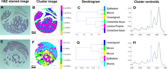
Biological materials presenting early signs of cancer would be beneficial for cancer screening/diagnosis. In this respect, the suitability of potentially exploiting mucus in colorectal cancer was tested using infrared spectroscopy in combination with statistical modeling. Twenty-six paraffinized colon tissue biopsy sections containing mucus regions from 20 individuals (10 normal and 16 cancerous) were measured using mid-infrared spectroscopic imaging. A digital de-paraffinization, followed by cluster analysis driven digital color-coded multi-staining segmented the infrared images into various histopathological features such as epithelium, connective tissue, stroma, and mucus regions within the tissue sections. Principal component analysis followed by supervised linear discriminant analysis was carried out on pure mucus and epithelial spectra from normal and cancerous regions of the tissue. For the mucus-based classification, a sensitivity of 96%, a specificity of 83%, and an area under the curve performance of 95% was obtained. For the epithelial tissue-based classification, a sensitivity of 72%, a specificity of 88%, and an area under the curve performance of 89% was obtained. The mucus spectral profiles further showed contributions indicative of glycans including that of sialic acid changes between these pathology groups. The study demonstrates that infrared spectroscopic analysis of mucus discriminates colorectal cancers with high sensitivity. This concept could be exploited to develop screening/diagnostic approaches complementary to histopathology.
中文翻译:
使用红外光谱表征结直肠粘液:肠癌筛查和诊断的潜在目标。
呈现癌症早期迹象的生物材料将有利于癌症筛查/诊断。在这方面,使用红外光谱结合统计模型测试了在结直肠癌中潜在利用粘液的适用性。使用中红外光谱成像测量了 26 个石蜡化结肠组织活检切片,其中包含来自 20 个个体(10 个正常和 16 个癌变)的粘液区域。数字脱蜡,然后进行聚类分析驱动的数字颜色编码多染色,将红外图像分割成各种组织病理学特征,例如组织切片内的上皮细胞、结缔组织、间质和粘液区域。对来自组织的正常和癌变区域的纯粘液和上皮光谱进行主成分分析,然后进行监督线性判别分析。对于基于粘液的分类,获得了 96% 的灵敏度、83% 的特异性和 95% 的曲线下面积。对于基于上皮组织的分类,获得了 72% 的灵敏度、88% 的特异性和 89% 的曲线下面积。粘液光谱图进一步显示了指示聚糖的贡献,包括这些病理组之间唾液酸变化的贡献。该研究表明,粘液的红外光谱分析能够以高灵敏度区分结直肠癌。这一概念可用于开发与组织病理学互补的筛查/诊断方法。


 京公网安备 11010802027423号