当前位置:
X-MOL 学术
›
ACS Appl. Nano Mater.
›
论文详情
Our official English website, www.x-mol.net, welcomes your feedback! (Note: you will need to create a separate account there.)
Synthesis of Ultrasmall Fe3O4 Nanoparticles as T1–T2 Dual-Modal Magnetic Resonance Imaging Contrast Agents in Rabbit Hepatic Tumors
ACS Applied Nano Materials ( IF 5.9 ) Pub Date : 2020-03-16 , DOI: 10.1021/acsanm.0c00306 Chen Bai 1 , Pengcheng Hu 2 , Nianlong Liu 3 , Guodong Feng 3 , Di Liu 1 , Yi Chen 1 , Ming Ma 1 , Ning Gu 1 , Yu Zhang 1
ACS Applied Nano Materials ( IF 5.9 ) Pub Date : 2020-03-16 , DOI: 10.1021/acsanm.0c00306 Chen Bai 1 , Pengcheng Hu 2 , Nianlong Liu 3 , Guodong Feng 3 , Di Liu 1 , Yi Chen 1 , Ming Ma 1 , Ning Gu 1 , Yu Zhang 1
Affiliation
|
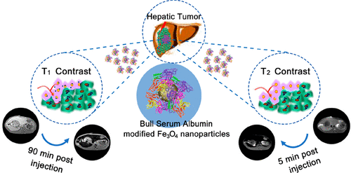
To elevate the accurate diagnosis of tumors, nanomaterial-based magnetic resonance imaging (MRI) contrast agents are being rapidly developed. In this paper, we report the large-scale synthetic method of Fe3O4 nanoparticles by high-temperature thermal decomposition and their application as time-dependent T1–T2 dual-modal MRI contrast agents in rabbit hepatic tumors. The Fe3O4 nanoparticles are modified with bull serum albumin (BSA), which provides excellent biocompatibility and colloidal stability. These nanoparticles also exhibit a high r1 value of 6.99 mM–1 s–1 and a low r2 value of 24.11 mM–1 s–1 at a clinically relevant magnetic field (3.0 T). In in vivo experiments, after intravenous administration with these nanoparticles, the rapid T2-weighted effect can be acquired within 5 min and the slow T1 contrast enhancement at the tumor site appears at 90 min. The explanation of this phenomenon is possibly due to the different accumulation rates and states of the nanoparticles in different tissues. In normal liver tissue, the nanoparticles can be quickly phagocytosed by a reticuloendothelial system and accumulated in the liver. These accumulated nanoparticles aggregate, improving the T2-weighted effect and reducing the T1-weighted effect. However, because of an enhanced permeation and retention effect, the nanoparticles are delivered into tumor tissue gradually and dispersedly, which takes a longer time to generate the T1-weighted effect at the tumor site. In summary, these Fe3O4 nanoparticles have significant potential to improve the diagnostic accuracy and sensitivity in MRI.
中文翻译:

T 1 - T 2双模态磁共振成像造影剂中超小Fe 3 O 4纳米颗粒在兔肝肿瘤中的合成
为了提高对肿瘤的准确诊断,基于纳米材料的磁共振成像(MRI)造影剂正在迅速开发中。在本文中,我们报道了通过高温热分解大规模合成Fe 3 O 4纳米颗粒的方法及其作为时间依赖性T 1 - T 2双峰MRI造影剂在兔肝肿瘤中的应用。Fe 3 O 4纳米颗粒经过牛血清白蛋白(BSA)修饰,具有出色的生物相容性和胶体稳定性。这些纳米颗粒还表现出6.99 mM –1 s –1的高r 1值。在临床相关磁场(3.0 T)下的r 2值为24.11 mM –1 s –1。在体内实验中,在静脉内给予这些纳米颗粒后,可以在5分钟内获得快速的T 2加权效应,而在肿瘤的90分钟处出现缓慢的T 1对比增强。这种现象的解释可能是由于纳米粒子在不同组织中的累积速率和状态不同所致。在正常肝组织中,纳米颗粒可以被网状内皮系统迅速吞噬并积累在肝脏中。这些累积的纳米颗粒聚集,改善了T 2加权效应和降低T 1加权效应。然而,由于增强的渗透和保留作用,纳米颗粒逐渐且分散地递送到肿瘤组织中,这花费更长的时间在肿瘤部位处产生T 1加权作用。总之,这些Fe 3 O 4纳米颗粒具有显着的潜力,可提高MRI的诊断准确性和灵敏度。
更新日期:2020-03-16
中文翻译:
T 1 - T 2双模态磁共振成像造影剂中超小Fe 3 O 4纳米颗粒在兔肝肿瘤中的合成
为了提高对肿瘤的准确诊断,基于纳米材料的磁共振成像(MRI)造影剂正在迅速开发中。在本文中,我们报道了通过高温热分解大规模合成Fe 3 O 4纳米颗粒的方法及其作为时间依赖性T 1 - T 2双峰MRI造影剂在兔肝肿瘤中的应用。Fe 3 O 4纳米颗粒经过牛血清白蛋白(BSA)修饰,具有出色的生物相容性和胶体稳定性。这些纳米颗粒还表现出6.99 mM –1 s –1的高r 1值。在临床相关磁场(3.0 T)下的r 2值为24.11 mM –1 s –1。在体内实验中,在静脉内给予这些纳米颗粒后,可以在5分钟内获得快速的T 2加权效应,而在肿瘤的90分钟处出现缓慢的T 1对比增强。这种现象的解释可能是由于纳米粒子在不同组织中的累积速率和状态不同所致。在正常肝组织中,纳米颗粒可以被网状内皮系统迅速吞噬并积累在肝脏中。这些累积的纳米颗粒聚集,改善了T 2加权效应和降低T 1加权效应。然而,由于增强的渗透和保留作用,纳米颗粒逐渐且分散地递送到肿瘤组织中,这花费更长的时间在肿瘤部位处产生T 1加权作用。总之,这些Fe 3 O 4纳米颗粒具有显着的潜力,可提高MRI的诊断准确性和灵敏度。


 京公网安备 11010802027423号