Nature Neuroscience ( IF 25.0 ) Pub Date : 2020-03-16 , DOI: 10.1038/s41593-020-0602-1 Omer Ali Bayraktar 1, 2, 3 , Theresa Bartels 1 , Staffan Holmqvist 1 , Vitalii Kleshchevnikov 3 , Araks Martirosyan 4 , Damon Polioudakis 5, 6 , Lucile Ben Haim 1 , Adam M H Young 7 , Mykhailo Y Batiuk 4 , Kirti Prakash 1 , Alexander Brown 8 , Kenny Roberts 3 , Mercedes F Paredes 2, 9 , Riki Kawaguchi 10 , John H Stockley 1 , Khalida Sabeur 1, 2 , Sandra M Chang 1, 2 , Eric Huang 9, 11 , Peter Hutchinson 7 , Erik M Ullian 12 , Martin Hemberg 3 , Giovanni Coppola 5, 10 , Matthew G Holt 4 , Daniel H Geschwind 5, 6 , David H Rowitch 1, 2
|
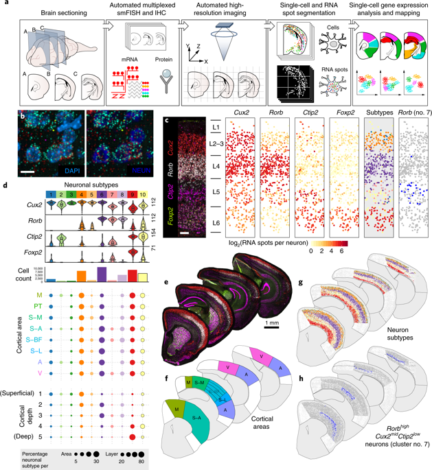
Although the cerebral cortex is organized into six excitatory neuronal layers, it is unclear whether glial cells show distinct layering. In the present study, we developed a high-content pipeline, the large-area spatial transcriptomic (LaST) map, which can quantify single-cell gene expression in situ. Screening 46 candidate genes for astrocyte diversity across the mouse cortex, we identified superficial, mid and deep astrocyte identities in gradient layer patterns that were distinct from those of neurons. Astrocyte layer features, established in the early postnatal cortex, mostly persisted in adult mouse and human cortex. Single-cell RNA sequencing and spatial reconstruction analysis further confirmed the presence of astrocyte layers in the adult cortex. Satb2 and Reeler mutations that shifted neuronal post-mitotic development were sufficient to alter glial layering, indicating an instructive role for neuronal cues. Finally, astrocyte layer patterns diverged between mouse cortical regions. These findings indicate that excitatory neurons and astrocytes are organized into distinct lineage-associated laminae.
中文翻译:
单细胞原位转录组图显示哺乳动物大脑皮层中的星形胶质细胞层。
尽管大脑皮层分为六个兴奋性神经元层,但尚不清楚神经胶质细胞是否表现出明显的分层。在本研究中,我们开发了一种高内涵管道,即大面积空间转录组(LaST)图谱,它可以原位量化单细胞基因表达。我们筛选了小鼠皮质中星形胶质细胞多样性的 46 个候选基因,确定了与神经元不同的梯度层模式的浅层、中层和深层星形胶质细胞特征。在出生后早期皮质中建立的星形胶质细胞层特征大部分持续存在于成年小鼠和人类皮质中。单细胞RNA测序和空间重建分析进一步证实了成人皮质中星形胶质细胞层的存在。Satb2和Reeler突变改变了神经元有丝分裂后的发育,足以改变神经胶质分层,表明神经元线索具有指导作用。最后,星形胶质细胞层模式在小鼠皮质区域之间存在差异。这些发现表明兴奋性神经元和星形胶质细胞被组织成不同的谱系相关层。


 京公网安备 11010802027423号