当前位置:
X-MOL 学术
›
Colloids Surf. B Biointerfaces
›
论文详情
Our official English website, www.x-mol.net, welcomes your feedback! (Note: you will need to create a separate account there.)
Effect of substrate topography on the regulation of human corneal stromal cells.
Colloids and Surfaces B: Biointerfaces ( IF 5.8 ) Pub Date : 2020-03-12 , DOI: 10.1016/j.colsurfb.2020.110971 Promita Bhattacharjee 1 , Brenton L Cavanagh 2 , Mark Ahearne 1
Colloids and Surfaces B: Biointerfaces ( IF 5.8 ) Pub Date : 2020-03-12 , DOI: 10.1016/j.colsurfb.2020.110971 Promita Bhattacharjee 1 , Brenton L Cavanagh 2 , Mark Ahearne 1
Affiliation
|
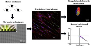
Optimal functionality of native corneal stroma depends on a well-ordered arrangement of extracellular matrix (ECM). To develop an in vitro corneal model, replication of the corneal in vivo microenvironment is needed. In this study, the impact of topographic cues on keratocyte phenotype is reported. Photolithography and polymer moulding were used to fabricate microgrooves on polydimethylsiloxane (PDMS) 2-2.5 μm deep and 5 μm, 10 μm, or 20 μm in width. Microgrooves constrained the cells body, compressed nuclei and led to cytoskeletal reorganization. It also influenced the concentration of actin filaments, condensation of chromatin and cell proliferation. Cells became more spread and actin filament concentration decreased as the microgroove width increased. Relationships were also demonstrated between microgroove width and cellular processes such as adhesion, migration and gene expression. Immunocytochemistry and gene expression (RT-PCR) analysis showed that microgroove width upregulated keratocyte specific genes. A microgroove with 5 μm width led to a pronounced alignment of cells along the edges of the microchannels and better supported cell polarization and migration compared with other microgroove widths or planar substrates. These findings provide important fundamental knowledge that could serve as a basis for better-controlled tissue growth and cell-engineering applications for corneal stroma regeneration through topographical patterns.
中文翻译:

底物形貌对人类角膜基质细胞调节的影响。
天然角膜基质的最佳功能取决于细胞外基质(ECM)的有序排列。为了建立体外角膜模型,需要在体内微环境复制角膜。在这项研究中,报道了地形线索对角化细胞表型的影响。光刻和聚合物成型用于在深度为2-2.5μm,宽度为5μm,10μm或20μm的聚二甲基硅氧烷(PDMS)上制造微沟槽。细槽限制了细胞体,压缩了细胞核并导致细胞骨架重组。它还影响肌动蛋白丝的浓度,染色质的凝聚和细胞增殖。随着微槽宽度的增加,细胞变得更加分散,肌动蛋白丝的浓度降低。还证明了微槽宽度与细胞过程如粘附,迁移和基因表达之间的关系。免疫细胞化学和基因表达(RT-PCR)分析表明,微槽宽度上调了角膜细胞特异性基因。与其他微沟槽宽度或平面基板相比,宽度为5μm的微沟槽导致细胞沿微通道边缘的明显对齐,并更好地支持了细胞的极化和迁移。这些发现提供了重要的基础知识,可作为通过地形图更好地控制组织生长和角膜基质再生的细胞工程应用的基础。免疫细胞化学和基因表达(RT-PCR)分析表明,微槽宽度上调了角膜细胞特异性基因。与其他微沟槽宽度或平面基板相比,宽度为5μm的微沟槽导致细胞沿微通道边缘的明显对齐,并更好地支持了细胞的极化和迁移。这些发现提供了重要的基础知识,可作为通过地形图更好地控制组织生长和角膜基质再生的细胞工程应用的基础。免疫细胞化学和基因表达(RT-PCR)分析表明,微槽宽度上调了角膜细胞特异性基因。与其他微沟槽宽度或平面基板相比,宽度为5μm的微沟槽导致细胞沿微通道边缘的明显对齐,并更好地支持了细胞的极化和迁移。这些发现提供了重要的基础知识,可作为通过地形图更好地控制组织生长和角膜基质再生的细胞工程应用的基础。
更新日期:2020-03-12
中文翻译:
底物形貌对人类角膜基质细胞调节的影响。
天然角膜基质的最佳功能取决于细胞外基质(ECM)的有序排列。为了建立体外角膜模型,需要在体内微环境复制角膜。在这项研究中,报道了地形线索对角化细胞表型的影响。光刻和聚合物成型用于在深度为2-2.5μm,宽度为5μm,10μm或20μm的聚二甲基硅氧烷(PDMS)上制造微沟槽。细槽限制了细胞体,压缩了细胞核并导致细胞骨架重组。它还影响肌动蛋白丝的浓度,染色质的凝聚和细胞增殖。随着微槽宽度的增加,细胞变得更加分散,肌动蛋白丝的浓度降低。还证明了微槽宽度与细胞过程如粘附,迁移和基因表达之间的关系。免疫细胞化学和基因表达(RT-PCR)分析表明,微槽宽度上调了角膜细胞特异性基因。与其他微沟槽宽度或平面基板相比,宽度为5μm的微沟槽导致细胞沿微通道边缘的明显对齐,并更好地支持了细胞的极化和迁移。这些发现提供了重要的基础知识,可作为通过地形图更好地控制组织生长和角膜基质再生的细胞工程应用的基础。免疫细胞化学和基因表达(RT-PCR)分析表明,微槽宽度上调了角膜细胞特异性基因。与其他微沟槽宽度或平面基板相比,宽度为5μm的微沟槽导致细胞沿微通道边缘的明显对齐,并更好地支持了细胞的极化和迁移。这些发现提供了重要的基础知识,可作为通过地形图更好地控制组织生长和角膜基质再生的细胞工程应用的基础。免疫细胞化学和基因表达(RT-PCR)分析表明,微槽宽度上调了角膜细胞特异性基因。与其他微沟槽宽度或平面基板相比,宽度为5μm的微沟槽导致细胞沿微通道边缘的明显对齐,并更好地支持了细胞的极化和迁移。这些发现提供了重要的基础知识,可作为通过地形图更好地控制组织生长和角膜基质再生的细胞工程应用的基础。


 京公网安备 11010802027423号