Nature Medicine ( IF 82.9 ) Pub Date : 2020-03-11 , DOI: 10.1038/s41591-020-0783-x Albert G Tsai 1 , David R Glass 1, 2 , Marisa Juntilla 1 , Felix J Hartmann 1 , Jean S Oak 1 , Sebastian Fernandez-Pol 1 , Robert S Ohgami 3 , Sean C Bendall 1, 2
|
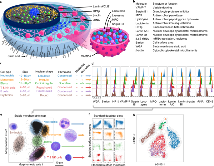
The diagnosis of lymphomas and leukemias requires hematopathologists to integrate microscopically visible cellular morphology with antibody-identified cell surface molecule expression. To merge these into one high-throughput, highly multiplexed, single-cell assay, we quantify cell morphological features by their underlying, antibody-measurable molecular components, which empowers mass cytometers to ‘see’ like pathologists. When applied to 71 diverse clinical samples, single-cell morphometric profiling reveals robust and distinct patterns of ‘morphometric’ markers for each major cell type. Individually, lamin B1 highlights acute leukemias, lamin A/C helps distinguish normal from neoplastic mature T cells, and VAMP-7 recapitulates light-cytometric side scatter. Combined with machine learning, morphometric markers form intuitive visualizations of normal and neoplastic cellular distribution and differentiation. When recalibrated for myelomonocytic blast enumeration, this approach is superior to flow cytometry and comparable to expert microscopy, bypassing years of specialized training. The contextualization of traditional surface markers on independent morphometric frameworks permits more sensitive and automated diagnosis of complex hematopoietic diseases.
中文翻译:
用于血液病理学诊断的多重单细胞形态测量
淋巴瘤和白血病的诊断需要血液病理学家将显微镜下可见的细胞形态与抗体识别的细胞表面分子表达相结合。为了将这些合并到一种高通量、高度多重的单细胞检测中,我们通过细胞的基础、抗体可测量的分子成分来量化细胞形态特征,这使得质谱流式细胞仪能够像病理学家一样“看到”。当应用于 71 个不同的临床样本时,单细胞形态测定分析揭示了每种主要细胞类型的稳健且独特的“形态测定”标记模式。单独而言,核纤层蛋白 B1 突出显示急性白血病,核纤层蛋白 A/C 有助于区分正常和肿瘤性成熟 T 细胞,VAMP-7 概括了光细胞侧向散射。与机器学习相结合,形态测量标记形成正常和肿瘤细胞分布和分化的直观可视化。当针对骨髓单核细胞原始计数进行重新校准时,这种方法优于流式细胞术,并且可与专家显微镜相媲美,无需多年的专业培训。传统表面标记在独立形态测量框架上的背景化允许对复杂的造血系统疾病进行更灵敏和自动化的诊断。


 京公网安备 11010802027423号