当前位置:
X-MOL 学术
›
J. Proteome Res.
›
论文详情
Our official English website, www.x-mol.net, welcomes your feedback! (Note: you will need to create a separate account there.)
Proteomic Profiling of Stem Cell Tissues during Regeneration of Deer Antler: A Model of Mammalian Organ Regeneration.
Journal of Proteome Research ( IF 4.4 ) Pub Date : 2020-03-10 , DOI: 10.1021/acs.jproteome.0c00026 Zhen Dong 1 , Stephen Haines 2 , Dawn Coates 1
Journal of Proteome Research ( IF 4.4 ) Pub Date : 2020-03-10 , DOI: 10.1021/acs.jproteome.0c00026 Zhen Dong 1 , Stephen Haines 2 , Dawn Coates 1
Affiliation
|
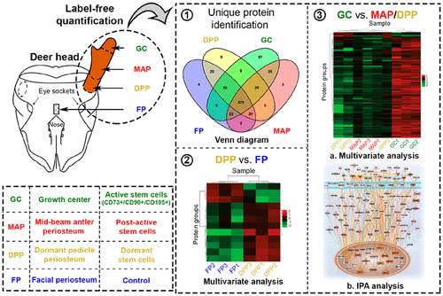
As the only known mammalian organ that can fully and annually regenerate, deer antler has significant advantages over lower-order animal models when investigating the control of stem-cell-based organ regeneration. Antler regeneration is known to be initiated and maintained by neural-crest-derived stem cells in different states of activation. Antler stem cells can therefore be used as a model to study proteins and pathways involved in the maintenance of a stem cell niche and their activation and differentiation during organ formation. In this study, the MSC markers CD73, CD90, and CD105 were examined within the antler tip. Label-free quantification was performed to investigate the protein profiles of antler stem cells under different stages of activation and included dormant pedicle periosteum (DPP), antler growth center (GC), post-active stem cells from mid-beam antler periosteum (MAP), and deer facial periosteum (FP) as a control (n = 3 per group). PEAKS and IPA software were used to analyze the proteomic data. Our research confirmed the central role of stem cell activation in the development of this mammalian organ by localizing the MSC markers within the antler growth center. Label-free quantification revealed that the greatest number of unique proteins (87) was found in the growth center. There were only 12 proteins found with expression levels that significantly differed between DPP and FP. Protein profiles of these two groups indicated that antler stem cells may use similar mechanisms to maintain dormancy within a stem cell niche. The number of significantly regulated proteins among DPP, MAP, and GC was 153. Among them, the majority were upregulated in the growth center. Activation of antler stem cells was associated with many biological processes and signaling pathways, such as Hippo and canonical Wnt signaling. This work identifies the key pathways, molecular/cellular functions, and upstream regulators involved in mammal organ regeneration. The mass spectrometry proteomics data have been deposited to the ProteomeXchange Consortium via the iProX partner repository with the dataset identifier PXD016824.
中文翻译:

鹿茸再生过程中干细胞组织的蛋白质组学分析:哺乳动物器官再生的模型。
鹿角是唯一已知的能够完全,每年再生的哺乳动物器官,在研究基于干细胞的器官再生的控制方法时,它相对于低阶动物模型具有明显的优势。已知鹿角的再生是由神经stem来源的干细胞在不同的激活状态下启动和维持的。因此,鹿茸干细胞可以用作研究蛋白质和干细胞生态位维持及其在器官形成过程中活化和分化的途径的模型。在这项研究中,在鹿角尖端内检查了MSC标记CD73,CD90和CD105。进行无标记定量研究在不同激活阶段的鹿角干细胞的蛋白质谱,包括休眠的椎弓根骨膜(DPP),鹿角生长中心(GC),ñ=每组3个)。PEAKS和IPA软件用于分析蛋白质组数据。我们的研究通过在鹿角生长中心定位MSC标记,证实了干细胞活化在该哺乳动物器官发育中的核心作用。无标记定量显示,在生长中心发现了最大数量的独特蛋白质(87)。仅发现12种蛋白,其表达水平在DPP和FP之间存在显着差异。这两类动物的蛋白质谱表明,鹿角干细胞可能使用类似的机制来维持干细胞生态位内的休眠。DPP,MAP和GC中受显着调节的蛋白质数量为153。其中,大多数在生长中心被上调。鹿角干细胞的激活与许多生物学过程和信号通路有关,例如河马和经典Wnt信号。这项工作确定了参与哺乳动物器官再生的关键途径,分子/细胞功能和上游调节剂。质谱蛋白质组学数据已通过iProX合作伙伴存储库以数据集标识符PXD016824存放到ProteomeXchange联盟。
更新日期:2020-04-24
中文翻译:
鹿茸再生过程中干细胞组织的蛋白质组学分析:哺乳动物器官再生的模型。
鹿角是唯一已知的能够完全,每年再生的哺乳动物器官,在研究基于干细胞的器官再生的控制方法时,它相对于低阶动物模型具有明显的优势。已知鹿角的再生是由神经stem来源的干细胞在不同的激活状态下启动和维持的。因此,鹿茸干细胞可以用作研究蛋白质和干细胞生态位维持及其在器官形成过程中活化和分化的途径的模型。在这项研究中,在鹿角尖端内检查了MSC标记CD73,CD90和CD105。进行无标记定量研究在不同激活阶段的鹿角干细胞的蛋白质谱,包括休眠的椎弓根骨膜(DPP),鹿角生长中心(GC),ñ=每组3个)。PEAKS和IPA软件用于分析蛋白质组数据。我们的研究通过在鹿角生长中心定位MSC标记,证实了干细胞活化在该哺乳动物器官发育中的核心作用。无标记定量显示,在生长中心发现了最大数量的独特蛋白质(87)。仅发现12种蛋白,其表达水平在DPP和FP之间存在显着差异。这两类动物的蛋白质谱表明,鹿角干细胞可能使用类似的机制来维持干细胞生态位内的休眠。DPP,MAP和GC中受显着调节的蛋白质数量为153。其中,大多数在生长中心被上调。鹿角干细胞的激活与许多生物学过程和信号通路有关,例如河马和经典Wnt信号。这项工作确定了参与哺乳动物器官再生的关键途径,分子/细胞功能和上游调节剂。质谱蛋白质组学数据已通过iProX合作伙伴存储库以数据集标识符PXD016824存放到ProteomeXchange联盟。


 京公网安备 11010802027423号