当前位置:
X-MOL 学术
›
Chem. Bio. Drug Des.
›
论文详情
Our official English website, www.x-mol.net, welcomes your feedback! (Note: you will need to create a separate account there.)
Design and synthesis of a novel peptide for selective detection of cancer cells.
Chemical Biology & Drug Design ( IF 3 ) Pub Date : 2020-03-09 , DOI: 10.1111/cbdd.13675 Krishnamoorthy Rajavenkatesh 1 , Murali Padmaja 2 , Indrakumar Janani 2 , Satish Aishwarya 2 , Korrapati Purna Sai 2 , Sathiah Thennarasu 1
Chemical Biology & Drug Design ( IF 3 ) Pub Date : 2020-03-09 , DOI: 10.1111/cbdd.13675 Krishnamoorthy Rajavenkatesh 1 , Murali Padmaja 2 , Indrakumar Janani 2 , Satish Aishwarya 2 , Korrapati Purna Sai 2 , Sathiah Thennarasu 1
Affiliation
|
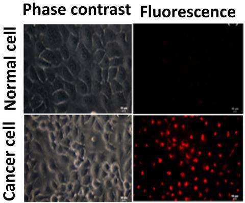
Using a minimalist approach, an 11‐residue peptide (Peptide 1 ) tagged with rhodamine fluorophore was designed and synthesized for selective detection of cancer cells. Peptide 1 contains RGD and NGR motifs to bind, respectively, integrins and aminopeptidase CD13, which are over expressed in cancer cells. Surface tension measurements revealed that peptide 1 possess surface‐active property owing to the overall hydrophobicity and cationic nature of the peptide. Peptide 1 displays cancer cell‐selective binding at ≤5.0 µM concentrations, while peptide 2 (randomized sequence of 1 ) shows non‐selective binding to normal and cancer cells. Fluorescence microscopy and FACS analysis demonstrated the intracellular localization of peptide 1 in three different cancer cell lines, confirming the role of RGD and NGR motifs. Cytotoxicity assay exhibited the viability of normal and cancer cells up to 100 µM concentrations of peptide 1 . Steady‐state fluorescence measurements disclosed the preferential interactions of the peptide 1 with anionic POPC/POPG bilayers rather than with zwitterionic POPC lipid bilayers. Circular dichroism studies showed minimal changes in the secondary structure of peptide 1 upon binding with the anionic lipid bilayers. Peptide 1 is largely unordered, non‐toxic, and useful for identification of cancer cells. Peptide 1 provides a template for designing drug‐loaded peptides for targeted delivery into cancer cells.
中文翻译:

设计和合成用于选择性检测癌细胞的新型肽。
使用极简方法,设计并合成了用罗丹明荧光团标记的11个残基的肽(肽1),用于选择性检测癌细胞。肽1含有RGD和NGR基序,分别结合在癌细胞中过度表达的整联蛋白和氨基肽酶CD13。表面张力测量结果表明,肽1具有由于总体疏水性和肽的阳离子性质的表面活性特性。肽1在≤5.0µM浓度下显示癌细胞选择性结合,而肽2(1的随机序列)显示与正常细胞和癌细胞的非选择性结合。荧光显微镜和FACS分析证明了肽1在三种不同癌细胞系中的细胞内定位,证实了RGD和NGR基序的作用。细胞毒性试验显示正常和癌细胞的活力高达100 µM肽1。稳态荧光测量揭示了肽1与阴离子POPC / POPG双层而不是两性离子POPC脂质双层的优先相互作用。圆二色性研究表明,与阴离子脂质双层结合后,肽1二级结构的变化很小。肽1基本上无序,无毒,可用于识别癌细胞。肽1提供了一个模板,用于设计可靶向递送至癌细胞的载药肽。
更新日期:2020-03-09
中文翻译:
设计和合成用于选择性检测癌细胞的新型肽。
使用极简方法,设计并合成了用罗丹明荧光团标记的11个残基的肽(肽1),用于选择性检测癌细胞。肽1含有RGD和NGR基序,分别结合在癌细胞中过度表达的整联蛋白和氨基肽酶CD13。表面张力测量结果表明,肽1具有由于总体疏水性和肽的阳离子性质的表面活性特性。肽1在≤5.0µM浓度下显示癌细胞选择性结合,而肽2(1的随机序列)显示与正常细胞和癌细胞的非选择性结合。荧光显微镜和FACS分析证明了肽1在三种不同癌细胞系中的细胞内定位,证实了RGD和NGR基序的作用。细胞毒性试验显示正常和癌细胞的活力高达100 µM肽1。稳态荧光测量揭示了肽1与阴离子POPC / POPG双层而不是两性离子POPC脂质双层的优先相互作用。圆二色性研究表明,与阴离子脂质双层结合后,肽1二级结构的变化很小。肽1基本上无序,无毒,可用于识别癌细胞。肽1提供了一个模板,用于设计可靶向递送至癌细胞的载药肽。


 京公网安备 11010802027423号