当前位置:
X-MOL 学术
›
Acta Biomater.
›
论文详情
Our official English website, www.x-mol.net, welcomes your feedback! (Note: you will need to create a separate account there.)
Advances in the Masquelet technique: Myeloid-derived suppressor cells promote angiogenesis in PMMA-induced membranes.
Acta Biomaterialia ( IF 9.7 ) Pub Date : 2020-03-09 , DOI: 10.1016/j.actbio.2020.03.010 Wenkai Wang 1 , Rui Zuo 1 , Haixia Long 2 , Yanqiu Wang 1 , Yang Zhang 1 , Chao Sun 1 , Gang Luo 1 , Yuan Zhang 1 , Changqing Li 1 , Yue Zhou 1 , Jie Li 1
Acta Biomaterialia ( IF 9.7 ) Pub Date : 2020-03-09 , DOI: 10.1016/j.actbio.2020.03.010 Wenkai Wang 1 , Rui Zuo 1 , Haixia Long 2 , Yanqiu Wang 1 , Yang Zhang 1 , Chao Sun 1 , Gang Luo 1 , Yuan Zhang 1 , Changqing Li 1 , Yue Zhou 1 , Jie Li 1
Affiliation
|
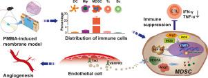
The periosteum plays a critical role in bone formation and defect reconstruction. The concept of tissue engineering in the periosteum has been suggested to solve the clinical problems related to bone defect repair. Insertion of polymethyl methacrylate (PMMA) bone cement can induce the autologous generation of a tissue-engineered periosteum and has been considered as a promising strategy for bone defect reconstruction. The PMMA-induced membrane is a crucial element in the reconstruction of bone defects, especially for angiogenesis, but its biological mechanism remains elusive. Here, a PMMA-induced membrane model was established using a femoral critically sized defect in mice. We identified myeloid-derived suppressor cells (MDSCs) as a regulatory component of induced membrane vascularization. The increased number of MDSCs was markedly linked to increased membrane thickness and capillary density. Importantly, the results of an in vitro coculture assay indicated that MDSCs of the induced membrane further facilitated the angiogenic capacity of human umbilical vein endothelial cells (HUVECs) by upregulating the expression of VEGFA, Ang2 and HIF-1α. Furthermore, signaling pathway blockade results suggested that STAT3 activation is involved in the upregulation of VEGFA, Ang2 and HIF-1α expression in induced membrane MDSCs. Our findings provide new insights into the mechanism of angiogenesis in the PMMA-induced membrane and confirm the key signaling molecules of MDSCs in induced membrane angiogenesis. Based on these results, this strategy may become a new therapy for the treatment of large bone defects in the future. STATEMENT OF SIGNIFICANCE: In this study, we established an autologous tissue-engineered periosteum - PMMA-induced membrane, which was formed by the foreign body reaction to PMMA bone cement. The induced membrane establishes a blood supply for the large bone defect healing. After investigation, our study discovered the critical cell type in the formation and angiogenesis processes of the induced membrane, myeloid-derived suppressor cells (MDSCs). We revealed that MDSCs of the induced membrane promote the angiogenesis of endothelial cells through the expression of VEGFA, Ang2 and HIF-1α, which was upregulated by the activation of STAT3 signaling. Our findings clarified the beneficial effect of MDSCs in the angiogenesis of bone repair, and offered an additional target for the study of foreign body reactions to bone repair materials.
中文翻译:

Masquelet技术的进展:髓样抑制细胞促进了PMMA诱导的膜中的血管生成。
骨膜在骨形成和缺损重建中起关键作用。已经提出了骨膜组织工程的概念来解决与骨缺损修复有关的临床问题。插入聚甲基丙烯酸甲酯(PMMA)骨水泥可以诱导组织工程性骨膜的自体生成,并被认为是骨缺损重建的一种有前途的策略。PMMA诱导的膜是重建骨缺损(尤其是血管生成)的关键元素,但其生物学机制仍然难以捉摸。在这里,使用小鼠的股骨临界大小缺陷建立了PMMA诱导的膜模型。我们确定了髓样抑制细胞(MDSCs)作为诱导膜血管化的调节成分。MDSC数量的增加与膜厚度和毛细管密度的增加显着相关。重要的是,体外共培养试验的结果表明,诱导膜的MDSC通过上调VEGFA,Ang2和HIF-1α的表达进一步促进人脐静脉内皮细胞(HUVEC)的血管生成能力。此外,信号通路的阻断结果表明,STAT3激活与诱导的膜MDSCs中VEGFA,Ang2和HIF-1α表达的上调有关。我们的发现为PMMA诱导的膜中的血管生成机制提供了新的见解,并确认了MDSCs在诱导的膜血管生成中的关键信号分子。基于这些结果,该策略将来可能成为治疗大骨缺损的一种新疗法。重大意义声明:在这项研究中,我们建立了一种自体组织工程化的骨膜-PMMA诱导膜,该膜是由异物与PMMA骨水泥反应形成的。诱导的膜为大骨缺损的愈合建立了血液供应。经过调查,我们的研究发现了诱导的膜,髓样抑制细胞(MDSCs)的形成和血管生成过程中的关键细胞类型。我们发现诱导膜的MDSCs通过VEGFA,Ang2和HIF-1α的表达促进内皮细胞的血管生成,而VEGFA,Ang2和HIF-1α的表达被STAT3信号的激活上调。我们的发现阐明了MDSCs在骨修复血管生成中的有益作用,并为研究异物对骨修复材料的反应提供了另一个目标。我们建立了自体组织工程化的骨膜-PMMA诱导的膜,该膜是由异物与PMMA骨水泥反应形成的。诱导的膜为大骨缺损的愈合建立了血液供应。经过调查,我们的研究发现了诱导的膜,髓样抑制细胞(MDSCs)的形成和血管生成过程中的关键细胞类型。我们发现诱导膜的MDSCs通过VEGFA,Ang2和HIF-1α的表达促进内皮细胞的血管生成,而VEGFA,Ang2和HIF-1α的表达被STAT3信号的激活上调。我们的发现阐明了MDSCs在骨修复血管生成中的有益作用,并为研究异物对骨修复材料的反应提供了另一个目标。我们建立了自体组织工程化的骨膜-PMMA诱导的膜,该膜是由异物与PMMA骨水泥反应形成的。诱导的膜为大骨缺损的愈合建立了血液供应。经过调查,我们的研究发现了诱导的膜,髓样抑制细胞(MDSCs)的形成和血管生成过程中的关键细胞类型。我们发现诱导膜的MDSCs通过VEGFA,Ang2和HIF-1α的表达促进内皮细胞的血管生成,而VEGFA,Ang2和HIF-1α的表达被STAT3信号的激活上调。我们的发现阐明了MDSCs在骨修复血管生成中的有益作用,并为研究异物对骨修复材料的反应提供了另一个目标。
更新日期:2020-03-09
中文翻译:
Masquelet技术的进展:髓样抑制细胞促进了PMMA诱导的膜中的血管生成。
骨膜在骨形成和缺损重建中起关键作用。已经提出了骨膜组织工程的概念来解决与骨缺损修复有关的临床问题。插入聚甲基丙烯酸甲酯(PMMA)骨水泥可以诱导组织工程性骨膜的自体生成,并被认为是骨缺损重建的一种有前途的策略。PMMA诱导的膜是重建骨缺损(尤其是血管生成)的关键元素,但其生物学机制仍然难以捉摸。在这里,使用小鼠的股骨临界大小缺陷建立了PMMA诱导的膜模型。我们确定了髓样抑制细胞(MDSCs)作为诱导膜血管化的调节成分。MDSC数量的增加与膜厚度和毛细管密度的增加显着相关。重要的是,体外共培养试验的结果表明,诱导膜的MDSC通过上调VEGFA,Ang2和HIF-1α的表达进一步促进人脐静脉内皮细胞(HUVEC)的血管生成能力。此外,信号通路的阻断结果表明,STAT3激活与诱导的膜MDSCs中VEGFA,Ang2和HIF-1α表达的上调有关。我们的发现为PMMA诱导的膜中的血管生成机制提供了新的见解,并确认了MDSCs在诱导的膜血管生成中的关键信号分子。基于这些结果,该策略将来可能成为治疗大骨缺损的一种新疗法。重大意义声明:在这项研究中,我们建立了一种自体组织工程化的骨膜-PMMA诱导膜,该膜是由异物与PMMA骨水泥反应形成的。诱导的膜为大骨缺损的愈合建立了血液供应。经过调查,我们的研究发现了诱导的膜,髓样抑制细胞(MDSCs)的形成和血管生成过程中的关键细胞类型。我们发现诱导膜的MDSCs通过VEGFA,Ang2和HIF-1α的表达促进内皮细胞的血管生成,而VEGFA,Ang2和HIF-1α的表达被STAT3信号的激活上调。我们的发现阐明了MDSCs在骨修复血管生成中的有益作用,并为研究异物对骨修复材料的反应提供了另一个目标。我们建立了自体组织工程化的骨膜-PMMA诱导的膜,该膜是由异物与PMMA骨水泥反应形成的。诱导的膜为大骨缺损的愈合建立了血液供应。经过调查,我们的研究发现了诱导的膜,髓样抑制细胞(MDSCs)的形成和血管生成过程中的关键细胞类型。我们发现诱导膜的MDSCs通过VEGFA,Ang2和HIF-1α的表达促进内皮细胞的血管生成,而VEGFA,Ang2和HIF-1α的表达被STAT3信号的激活上调。我们的发现阐明了MDSCs在骨修复血管生成中的有益作用,并为研究异物对骨修复材料的反应提供了另一个目标。我们建立了自体组织工程化的骨膜-PMMA诱导的膜,该膜是由异物与PMMA骨水泥反应形成的。诱导的膜为大骨缺损的愈合建立了血液供应。经过调查,我们的研究发现了诱导的膜,髓样抑制细胞(MDSCs)的形成和血管生成过程中的关键细胞类型。我们发现诱导膜的MDSCs通过VEGFA,Ang2和HIF-1α的表达促进内皮细胞的血管生成,而VEGFA,Ang2和HIF-1α的表达被STAT3信号的激活上调。我们的发现阐明了MDSCs在骨修复血管生成中的有益作用,并为研究异物对骨修复材料的反应提供了另一个目标。


 京公网安备 11010802027423号