当前位置:
X-MOL 学术
›
ACS Macro Lett.
›
论文详情
Our official English website, www.x-mol.net, welcomes your feedback! (Note: you will need to create a separate account there.)
Facile Dye-Initiated Polymerization of Lactide–Glycolide Generates Highly Fluorescent Poly(lactic-co-glycolic Acid) for Enhanced Characterization of Cellular Delivery
ACS Macro Letters ( IF 5.8 ) Pub Date : 2020-03-06 , DOI: 10.1021/acsmacrolett.9b01014 Mohammad A Al-Natour 1, 2, 3 , Mohamed D Yousif 1 , Robert Cavanagh 1 , Amjad Abouselo 1 , Edward A Apebende 4 , Amir Ghaemmaghami 2 , Dong-Hyun Kim 1 , Jonathan W Aylott 1 , Vincenzo Taresco 1, 4 , Veeren M Chauhan 1 , Cameron Alexander 1
ACS Macro Letters ( IF 5.8 ) Pub Date : 2020-03-06 , DOI: 10.1021/acsmacrolett.9b01014 Mohammad A Al-Natour 1, 2, 3 , Mohamed D Yousif 1 , Robert Cavanagh 1 , Amjad Abouselo 1 , Edward A Apebende 4 , Amir Ghaemmaghami 2 , Dong-Hyun Kim 1 , Jonathan W Aylott 1 , Vincenzo Taresco 1, 4 , Veeren M Chauhan 1 , Cameron Alexander 1
Affiliation
|
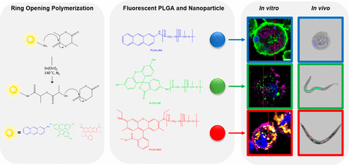
Poly(lactic-co-glycolic acid) (PLGA) is a versatile synthetic copolymer that is widely used in pharmaceutical applications. This is because it is well-tolerated in the body, and copolymers of varying physicochemical properties are readily available via ring-opening polymerization. However, native PLGA polymers are hard to track as drug delivery carriers when delivered to subcellular spaces, due to the absence of an easily accessible “handle” for fluorescent labeling. Here we show a one-step, scalable, solvent-free, synthetic route to fluorescent blue (2-aminoanthracene), green (5-aminofluorescein), and red (rhodamine-6G) PLGA, in which every polymer chain in the sample is fluorescently labeled. The utility of initiator-labeled PLGA was demonstrated through the preparation of nanoparticles, capable of therapeutic subcellular delivery to T-helper-precursor-1 (THP-1) macrophages, a model cell line for determining in vitro biocompatibility and particle uptake. Super resolution confocal fluorescence microscopy imaging showed that dye-initiated PLGA nanoparticles were internalized to punctate regions and retained bright fluorescence over at least 24 h. In comparison, PLGA nanoparticles with 5-aminofluorescein introduced by conventional nanoprecipitation/encapsulation showed diffuse and much lower fluorescence intensity in the same cells and over the same time periods. The utility of this approach for in vitro drug delivery experiments was demonstrated through the concurrent imaging of the fluorescent drug doxorubicin (λex = 480 nm, λem = 590 nm) with carrier 5-aminofluorescein PLGA, also in THP-1 cells, in which the intracellular locations of the drug and the polymer could be clearly visualized. Finally, the dye-labeled particles were evaluated in an in vivo model, via delivery to the nematode Caenorhabditis elegans, with bright fluorescence again apparent in the internal tract after 3 h. The results presented in this manuscript highlight the ease of synthesis of highly fluorescent PLGA, which could be used to augment tracking of future therapeutics and accelerate in vitro and in vivo characterization of delivery systems prior to clinical translation.
中文翻译:

染料引发的丙交酯-乙交酯聚合反应生成高荧光聚(乳酸-共-乙醇酸)以增强细胞递送的表征
聚乳酸-乙醇酸) (PLGA) 是一种多功能合成共聚物,广泛用于制药应用。这是因为它在体内具有良好的耐受性,并且通过开环聚合很容易获得具有不同物理化学性质的共聚物。然而,天然 PLGA 聚合物在递送到亚细胞空间时很难作为药物递送载体进行追踪,因为缺乏用于荧光标记的易于访问的“手柄”。在这里,我们展示了一种生成荧光蓝(2-氨基蒽)、绿色(5-氨基荧光素)和红色(罗丹明-6G)PLGA 的一步、可扩展、无溶剂合成路线,其中样品中的每个聚合物链都是荧光标记。通过制备纳米颗粒证明了引发剂标记的 PLGA 的效用,体外生物相容性和颗粒吸收。超分辨率共聚焦荧光显微镜成像显示染料引发的 PLGA 纳米粒子被内化为点状区域并在至少 24 小时内保持明亮的荧光。相比之下,通过常规纳米沉淀/封装引入的具有 5-氨基荧光素的 PLGA 纳米粒子在相同的细胞中和相同的时间段内显示出弥散性和低得多的荧光强度。通过对荧光药物阿霉素(λ ex = 480 nm,λ em= 590 nm)与载体 5-氨基荧光素 PLGA,也在 THP-1 细胞中,其中药物和聚合物的细胞内位置可以清楚地看到。最后,在体内模型中对染料标记的颗粒进行了评估,通过递送至秀丽隐杆线虫,3 小时后内部通道再次出现明亮的荧光。本手稿中的结果突出了高荧光 PLGA 合成的简易性,可用于增强对未来治疗的跟踪,并在临床转化之前加速递送系统的体外和体内表征。
更新日期:2020-03-06
中文翻译:
染料引发的丙交酯-乙交酯聚合反应生成高荧光聚(乳酸-共-乙醇酸)以增强细胞递送的表征
聚乳酸-乙醇酸) (PLGA) 是一种多功能合成共聚物,广泛用于制药应用。这是因为它在体内具有良好的耐受性,并且通过开环聚合很容易获得具有不同物理化学性质的共聚物。然而,天然 PLGA 聚合物在递送到亚细胞空间时很难作为药物递送载体进行追踪,因为缺乏用于荧光标记的易于访问的“手柄”。在这里,我们展示了一种生成荧光蓝(2-氨基蒽)、绿色(5-氨基荧光素)和红色(罗丹明-6G)PLGA 的一步、可扩展、无溶剂合成路线,其中样品中的每个聚合物链都是荧光标记。通过制备纳米颗粒证明了引发剂标记的 PLGA 的效用,体外生物相容性和颗粒吸收。超分辨率共聚焦荧光显微镜成像显示染料引发的 PLGA 纳米粒子被内化为点状区域并在至少 24 小时内保持明亮的荧光。相比之下,通过常规纳米沉淀/封装引入的具有 5-氨基荧光素的 PLGA 纳米粒子在相同的细胞中和相同的时间段内显示出弥散性和低得多的荧光强度。通过对荧光药物阿霉素(λ ex = 480 nm,λ em= 590 nm)与载体 5-氨基荧光素 PLGA,也在 THP-1 细胞中,其中药物和聚合物的细胞内位置可以清楚地看到。最后,在体内模型中对染料标记的颗粒进行了评估,通过递送至秀丽隐杆线虫,3 小时后内部通道再次出现明亮的荧光。本手稿中的结果突出了高荧光 PLGA 合成的简易性,可用于增强对未来治疗的跟踪,并在临床转化之前加速递送系统的体外和体内表征。


 京公网安备 11010802027423号