当前位置:
X-MOL 学术
›
J. Hepatol.
›
论文详情
Our official English website, www.x-mol.net, welcomes your feedback! (Note: you will need to create a separate account there.)
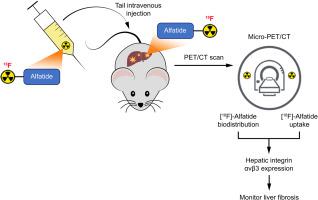
[18F]- Alfatide PET imaging of integrin αvβ3 for the non-invasive quantification of liver fibrosis
Journal of Hepatology ( IF 25.7 ) Pub Date : 2020-07-01 , DOI: 10.1016/j.jhep.2020.02.018 Tuo Shao 1 , Zhen Chen 2 , Vasily Belov 3 , Xiaohong Wang 4 , Steve H Rwema 5 , Viksit Kumar 4 , Hualong Fu 2 , Xiaoyun Deng 2 , Jian Rong 2 , Qingzhen Yu 2 , Lixin Lang 6 , Wenyu Lin 5 , Lee Josephson 2 , Anthony E Samir 4 , Xiaoyuan Chen 6 , Raymond T Chung 5 , Steven H Liang 2
Journal of Hepatology ( IF 25.7 ) Pub Date : 2020-07-01 , DOI: 10.1016/j.jhep.2020.02.018 Tuo Shao 1 , Zhen Chen 2 , Vasily Belov 3 , Xiaohong Wang 4 , Steve H Rwema 5 , Viksit Kumar 4 , Hualong Fu 2 , Xiaoyun Deng 2 , Jian Rong 2 , Qingzhen Yu 2 , Lixin Lang 6 , Wenyu Lin 5 , Lee Josephson 2 , Anthony E Samir 4 , Xiaoyuan Chen 6 , Raymond T Chung 5 , Steven H Liang 2
Affiliation
|
BACKGROUND & AIMS
The vitronectin receptor integrin alpha v beta 3 (αvβ3) drives fibrogenic activation of hepatic stellate cells (HSC). Molecular imaging targeting the integrin αvβ3 could provide a non-invasive method for evaluating the expression and the function of the integrin αvβ3 on activated HSCs (aHSCs) in the injured liver. In this study, we sought to compare differences in uptake of the [18F]-Alfatide between normal and injured liver to evaluate its utility for assessment of hepatic fibrogenesis. METHODS
PET with [18F]-Alfatide, non-enhanced computerized tomography (CT), histopathology, immunofluorescence staining, immunoblotting and gene analysis were performed to evaluate and quantify hepatic integrin αvβ3 levels and liver fibrosis progression in carbon-tetrachloride (CCl4) and bile duct ligation (BDL) induced liver fibrosis mice model. The AUC liver 0-30 min to AUC blood 0-30 min contrast was used as an integrin αvβ3-PET index to quantify fibrosis progression. Ex vivo analysis of frozen liver tissue from patients with fibrosis and cirrhosis verified the animal findings. RESULTS
Fibrotic mouse livers showed enhanced [18F]-Alfatide uptake and retention compared to control livers. The radiotracer was demonstrated to bind specifically with integrin αvβ3 mainly expressed on aHSCs. Autoradiography and histopathology confirmed the PET imaging results. Further, the mRNA and protein level of integrin αvβ3 and its signaling complex were higher in CCl4 and BDL models compared to controls. Human fibrotic liver section results supported the animal findings. CONCLUSIONS
Imaging hepatic integrin αvβ3 with PET and [18F]-Alfatide offers a potential noninvasive method for monitoring the progression of liver fibrosis.
中文翻译:

[18F]- 整合素 αvβ3 的 Alfatide PET 成像用于肝纤维化的非侵入性定量
背景和目的玻连蛋白受体整合素α v β 3 (αvβ3) 驱动肝星状细胞(HSC) 的纤维化激活。靶向整合素 αvβ3 的分子成像可以提供一种非侵入性方法来评估整合素 αvβ3 在受损肝脏中活化的 HSC (aHSC) 上的表达和功能。在这项研究中,我们试图比较正常肝脏和受损肝脏之间 [18F]-Alfatide 摄取的差异,以评估其在评估肝纤维化方面的效用。方法 PET 与 [18F]-Alfatide、非增强计算机断层扫描 (CT)、组织病理学、免疫荧光染色、进行免疫印迹和基因分析以评估和量化四氯化碳 (CCl4) 和胆管结扎 (BDL) 诱导的肝纤维化小鼠模型中的肝整合素 αvβ3 水平和肝纤维化进展。AUC 肝脏 0-30 分钟与 AUC 血液 0-30 分钟对比被用作整合素αvβ3-PET 指数以量化纤维化进展。对来自纤维化和肝硬化患者的冷冻肝组织的离体分析证实了动物的发现。结果 与对照肝脏相比,纤维化小鼠肝脏显示出增强的 [18F]-Alfatide 摄取和保留。已证明放射性示踪剂与主要在 aHSC 上表达的整合素 αvβ3 特异性结合。放射自显影和组织病理学证实了 PET 成像结果。更远,与对照相比,CCl4 和 BDL 模型中整联蛋白 αvβ3 及其信号复合物的 mRNA 和蛋白质水平更高。人类纤维化肝切片结果支持动物研究结果。结论 使用 PET 和 [18F]-Alfatide 对肝整联蛋白 αvβ3 进行成像为监测肝纤维化进展提供了一种潜在的无创方法。
更新日期:2020-07-01
中文翻译:
[18F]- 整合素 αvβ3 的 Alfatide PET 成像用于肝纤维化的非侵入性定量
背景和目的玻连蛋白受体整合素α v β 3 (αvβ3) 驱动肝星状细胞(HSC) 的纤维化激活。靶向整合素 αvβ3 的分子成像可以提供一种非侵入性方法来评估整合素 αvβ3 在受损肝脏中活化的 HSC (aHSC) 上的表达和功能。在这项研究中,我们试图比较正常肝脏和受损肝脏之间 [18F]-Alfatide 摄取的差异,以评估其在评估肝纤维化方面的效用。方法 PET 与 [18F]-Alfatide、非增强计算机断层扫描 (CT)、组织病理学、免疫荧光染色、进行免疫印迹和基因分析以评估和量化四氯化碳 (CCl4) 和胆管结扎 (BDL) 诱导的肝纤维化小鼠模型中的肝整合素 αvβ3 水平和肝纤维化进展。AUC 肝脏 0-30 分钟与 AUC 血液 0-30 分钟对比被用作整合素αvβ3-PET 指数以量化纤维化进展。对来自纤维化和肝硬化患者的冷冻肝组织的离体分析证实了动物的发现。结果 与对照肝脏相比,纤维化小鼠肝脏显示出增强的 [18F]-Alfatide 摄取和保留。已证明放射性示踪剂与主要在 aHSC 上表达的整合素 αvβ3 特异性结合。放射自显影和组织病理学证实了 PET 成像结果。更远,与对照相比,CCl4 和 BDL 模型中整联蛋白 αvβ3 及其信号复合物的 mRNA 和蛋白质水平更高。人类纤维化肝切片结果支持动物研究结果。结论 使用 PET 和 [18F]-Alfatide 对肝整联蛋白 αvβ3 进行成像为监测肝纤维化进展提供了一种潜在的无创方法。


 京公网安备 11010802027423号