当前位置:
X-MOL 学术
›
Microsc. Res. Tech.
›
论文详情
Our official English website, www.x-mol.net, welcomes your feedback! (Note: you will need to create a separate account there.)
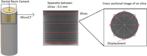
Microporosity and polymerization contraction as function of depth in dental resin cements by X-ray computed microtomography.
Microscopy Research and Technique ( IF 2.5 ) Pub Date : 2020-03-02 , DOI: 10.1002/jemt.23456 Rafaela Gheller 1 , Adrieli Burey 1 , Bruno Luiz Santana Vicentin 2 , Paulo José Dos Reis 3 , Carlos Roberto Appoloni 2 , Cássia Cilene Dezan Garbelini 4 , Márcio Grama Hoeppner 1
Microscopy Research and Technique ( IF 2.5 ) Pub Date : 2020-03-02 , DOI: 10.1002/jemt.23456 Rafaela Gheller 1 , Adrieli Burey 1 , Bruno Luiz Santana Vicentin 2 , Paulo José Dos Reis 3 , Carlos Roberto Appoloni 2 , Cássia Cilene Dezan Garbelini 4 , Márcio Grama Hoeppner 1
Affiliation
|
This research aimed to obtain the depth dependence of polymerization contraction and microporosity from irradiated dental resin cements by X‐ray computed microtomography (μCT). Samples (n = 5) of commercial Relyx U200 (RU) and AllCem Core (AC) dual‐cure resin cements were injected in a cylindrical Teflon sampler (25 mm3) and separated according to polymerization mechanism: self‐cured (not irradiated) and dual‐cured (irradiated from the top surface with a LED device). The cement's volume was scanned with the μCT scanning conditions kept constant. To assess the depth dependence of polymerization contraction, it was measured the displacement of the cement mass from the sample holder at 30 vertical cuts (0.1 mm distant). To probe the microporosity, the percentage of area with presence of porosity by slice was obtained. All data were statistically treated. It was observed a positive linear correlation between depth and polymerization contraction in the irradiated groups. In the other hand, the concentration of micropores decreased with increasing depth. Furthermore, the composition of the resin cement was determinant for the correlation's coefficients of these physical properties with depth. The μCT technique showed to be useful to probe physical properties of dental restorative materials that influence in the clinical outcomes, revealing that, for thin specimens, when light cured the RU cement presented mechanical behavior more favorable for clinical applications.
中文翻译:

通过X射线计算机显微断层照相术,牙科树脂胶结物中的微孔性和聚合收缩随深度变化。
这项研究旨在通过X射线计算机显微断层扫描(μCT)从辐照的牙科树脂胶结物中获得聚合收缩和微孔率的深度依赖性。将商用Relyx U200(RU)和AllCem Core(AC)双固化树脂水泥样品(n = 5)注入到圆柱铁氟龙采样器(25 mm 3)并根据聚合机理进行分离:自固化(未辐照)和双固化(通过LED器件从顶面辐照)。在μCT扫描条件保持不变的情况下扫描水泥的体积。为了评估聚合收缩的深度依赖性,在30个垂直切口(相距0.1毫米)处测量了水泥块从样品架上的位移。为了探测微孔,通过切片获得存在孔隙的面积的百分比。所有数据均经过统计学处理。观察到在照射组中深度与聚合收缩之间呈线性正相关。另一方面,微孔的浓度随着深度的增加而降低。此外,树脂水泥的组成是相关性的决定因素。这些物理性质的系数随深度变化。μCT技术被证明可用于探测影响临床结果的牙科修复材料的物理性能,从而揭示出,对于薄样品,光固化后的RU水泥呈现出对临床应用更有利的机械性能。
更新日期:2020-03-02
中文翻译:
通过X射线计算机显微断层照相术,牙科树脂胶结物中的微孔性和聚合收缩随深度变化。
这项研究旨在通过X射线计算机显微断层扫描(μCT)从辐照的牙科树脂胶结物中获得聚合收缩和微孔率的深度依赖性。将商用Relyx U200(RU)和AllCem Core(AC)双固化树脂水泥样品(n = 5)注入到圆柱铁氟龙采样器(25 mm 3)并根据聚合机理进行分离:自固化(未辐照)和双固化(通过LED器件从顶面辐照)。在μCT扫描条件保持不变的情况下扫描水泥的体积。为了评估聚合收缩的深度依赖性,在30个垂直切口(相距0.1毫米)处测量了水泥块从样品架上的位移。为了探测微孔,通过切片获得存在孔隙的面积的百分比。所有数据均经过统计学处理。观察到在照射组中深度与聚合收缩之间呈线性正相关。另一方面,微孔的浓度随着深度的增加而降低。此外,树脂水泥的组成是相关性的决定因素。这些物理性质的系数随深度变化。μCT技术被证明可用于探测影响临床结果的牙科修复材料的物理性能,从而揭示出,对于薄样品,光固化后的RU水泥呈现出对临床应用更有利的机械性能。


 京公网安备 11010802027423号