当前位置:
X-MOL 学术
›
Microsc. Res. Tech.
›
论文详情
Our official English website, www.x-mol.net, welcomes your feedback! (Note: you will need to create a separate account there.)
Morphological and functional characterization of circulating hemocytes using microscopy techniques.
Microscopy Research and Technique ( IF 2.5 ) Pub Date : 2020-02-28 , DOI: 10.1002/jemt.23463 Sivakamavalli Jeyachandran 1, 2 , Kiyun Park 1 , Ihn-Sil Kwak 1, 3 , Vaseeharan Baskaralingam 2
Microscopy Research and Technique ( IF 2.5 ) Pub Date : 2020-02-28 , DOI: 10.1002/jemt.23463 Sivakamavalli Jeyachandran 1, 2 , Kiyun Park 1 , Ihn-Sil Kwak 1, 3 , Vaseeharan Baskaralingam 2
Affiliation
|
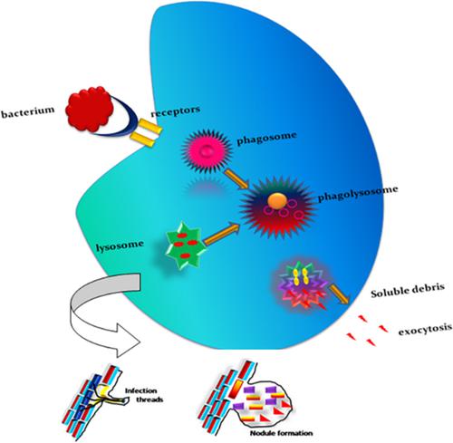
In the present study, Microscopy studies were performed to characterize the blood cells of the mangrove crab Episesarma tetragonum. Three types of hemocytes were observed: granulocytes, semi‐granulocytes, and hyalinocytes or agranulocytes. Hyalinocytes have a distinguished nucleus surrounded by the cytoplasm, and a peculiar cell type was present throughout the cytosol, lysosomes with hemocyte types (granules) stained red (pink). Giemsa staining was used to differentiate between the large and small hemocytes. Ehrlich's staining was used to differentiate granule‐containing cells in acidophils (55%), basophils (44%), and neutrophils (<1%). Periodic acid–Schiff staining was used to identify the sugar molecules in the cytoplasm. Cell‐mediated immune reactions including phagocytosis, encapsulation, agglutination, and peroxidase‐mediated cell adhesion are the functions of hemocytes. Agglutination reaction involves both kind of cells involved in yeast and heme‐agglutination responses in invertebrates. The beta glucan outer layer of yeast cells was recognized by hemocyte receptors. Human RBC cells were agglutinated via granulocytes. E. tetragonum hemocytes are an important animal model for studying both ultrastructural and functional activity of circulating cells. In addition, E. tetragonum hemocytes exhibited excellent antibacterial and antibiofilm activities were studied through plating and microplate assays. Biofilm inhibition was also visualized through changes in biochemical assays and morphological variations were visualized through levels in in situ microscopy analysis.
中文翻译:

使用显微镜技术对循环血细胞的形态和功能进行表征。
在本研究中,进行了显微镜研究以鉴定红树林蟹Episesarma tetragonum的血细胞。观察到三种类型的血细胞:粒细胞,半粒细胞和透明质细胞或粒细胞。透明质细胞具有被细胞质包围的独特核,并且在整个细胞质中存在特殊的细胞类型,溶血体的血细胞类型(颗粒)被染成红色(粉红色)。Giemsa染色用于区分大血细胞和小血细胞。Ehrlich染色用于区分嗜酸细胞(55%),嗜碱细胞(44%)和嗜中性粒细胞(<1%)中的含颗粒细胞。高碘酸-席夫氏染色用于鉴定细胞质中的糖分子。细胞介导的免疫反应包括吞噬作用,包囊,凝集和过氧化物酶介导的细胞粘附是血细胞的功能。凝集反应既涉及酵母中涉及的细胞类型,也涉及无脊椎动物中的血红素凝集反应。酵母细胞的β-葡聚糖外层被血细胞受体识别。通过粒细胞凝集人RBC细胞。四角马血红细胞是研究循环细胞超微结构和功能活性的重要动物模型。此外,通过平板接种和微孔板检测研究了四角马血红细胞具有优异的抗菌和抗生物膜活性。生物膜抑制作用还可以通过生化测定法的变化来观察,形态变化可以通过原位显微镜分析的水平来观察。
更新日期:2020-02-28
中文翻译:
使用显微镜技术对循环血细胞的形态和功能进行表征。
在本研究中,进行了显微镜研究以鉴定红树林蟹Episesarma tetragonum的血细胞。观察到三种类型的血细胞:粒细胞,半粒细胞和透明质细胞或粒细胞。透明质细胞具有被细胞质包围的独特核,并且在整个细胞质中存在特殊的细胞类型,溶血体的血细胞类型(颗粒)被染成红色(粉红色)。Giemsa染色用于区分大血细胞和小血细胞。Ehrlich染色用于区分嗜酸细胞(55%),嗜碱细胞(44%)和嗜中性粒细胞(<1%)中的含颗粒细胞。高碘酸-席夫氏染色用于鉴定细胞质中的糖分子。细胞介导的免疫反应包括吞噬作用,包囊,凝集和过氧化物酶介导的细胞粘附是血细胞的功能。凝集反应既涉及酵母中涉及的细胞类型,也涉及无脊椎动物中的血红素凝集反应。酵母细胞的β-葡聚糖外层被血细胞受体识别。通过粒细胞凝集人RBC细胞。四角马血红细胞是研究循环细胞超微结构和功能活性的重要动物模型。此外,通过平板接种和微孔板检测研究了四角马血红细胞具有优异的抗菌和抗生物膜活性。生物膜抑制作用还可以通过生化测定法的变化来观察,形态变化可以通过原位显微镜分析的水平来观察。


 京公网安备 11010802027423号