当前位置:
X-MOL 学术
›
J. Intern. Med.
›
论文详情
Our official English website, www.x-mol.net, welcomes your feedback! (Note: you will need to create a separate account there.)
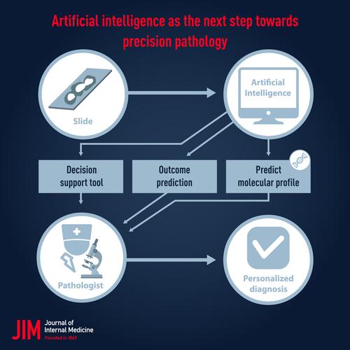
Artificial intelligence as the next step towards precision pathology.
Journal of Internal Medicine ( IF 11.1 ) Pub Date : 2020-03-03 , DOI: 10.1111/joim.13030 B Acs 1 , M Rantalainen 2 , J Hartman 1
Journal of Internal Medicine ( IF 11.1 ) Pub Date : 2020-03-03 , DOI: 10.1111/joim.13030 B Acs 1 , M Rantalainen 2 , J Hartman 1
Affiliation
|
Pathology is the cornerstone of cancer care. The need for accuracy in histopathologic diagnosis of cancer is increasing as personalized cancer therapy requires accurate biomarker assessment. The appearance of digital image analysis holds promise to improve both the volume and precision of histomorphological evaluation. Recently, machine learning, and particularly deep learning, has enabled rapid advances in computational pathology. The integration of machine learning into routine care will be a milestone for the healthcare sector in the next decade, and histopathology is right at the centre of this revolution. Examples of potential high‐value machine learning applications include both model‐based assessment of routine diagnostic features in pathology, and the ability to extract and identify novel features that provide insights into a disease. Recent groundbreaking results have demonstrated that applications of machine learning methods in pathology significantly improves metastases detection in lymph nodes, Ki67 scoring in breast cancer, Gleason grading in prostate cancer and tumour‐infiltrating lymphocyte (TIL) scoring in melanoma. Furthermore, deep learning models have also been demonstrated to be able to predict status of some molecular markers in lung, prostate, gastric and colorectal cancer based on standard HE slides. Moreover, prognostic (survival outcomes) deep neural network models based on digitized HE slides have been demonstrated in several diseases, including lung cancer, melanoma and glioma. In this review, we aim to present and summarize the latest developments in digital image analysis and in the application of artificial intelligence in diagnostic pathology.
中文翻译:

人工智能是走向精确病理学的下一步。
病理学是癌症治疗的基石。随着个性化癌症治疗需要准确的生物标志物评估,对癌症的组织病理学诊断的准确性的需求正在增加。数字图像分析的出现有望改善组织形态学评估的数量和准确性。最近,机器学习,尤其是深度学习,已经使计算病理学迅速发展。将机器学习整合到常规护理中将是未来十年医疗保健行业的一个里程碑,而组织病理学正是这场革命的核心。潜在的高价值机器学习应用程序的示例包括对病理学中常规诊断特征进行基于模型的评估,以及提取和识别新颖特征以了解疾病的能力。最新的开创性结果表明,机器学习方法在病理学中的应用显着改善了淋巴结转移检测,乳腺癌的Ki67评分,前列腺癌的Gleason评分以及黑素瘤的肿瘤浸润淋巴细胞(TIL)评分。此外,还已经证明深度学习模型能够基于标准HE幻灯片预测肺癌,前列腺癌,胃癌和结肠直肠癌中某些分子标记的状态。此外,已经在多种疾病(包括肺癌,黑色素瘤和神经胶质瘤)中证明了基于数字化HE幻灯片的预后(生存结果)深层神经网络模型。在这篇综述中,我们旨在介绍和总结数字图像分析以及人工智能在诊断病理学中的最新发展。
更新日期:2020-03-03
中文翻译:
人工智能是走向精确病理学的下一步。
病理学是癌症治疗的基石。随着个性化癌症治疗需要准确的生物标志物评估,对癌症的组织病理学诊断的准确性的需求正在增加。数字图像分析的出现有望改善组织形态学评估的数量和准确性。最近,机器学习,尤其是深度学习,已经使计算病理学迅速发展。将机器学习整合到常规护理中将是未来十年医疗保健行业的一个里程碑,而组织病理学正是这场革命的核心。潜在的高价值机器学习应用程序的示例包括对病理学中常规诊断特征进行基于模型的评估,以及提取和识别新颖特征以了解疾病的能力。最新的开创性结果表明,机器学习方法在病理学中的应用显着改善了淋巴结转移检测,乳腺癌的Ki67评分,前列腺癌的Gleason评分以及黑素瘤的肿瘤浸润淋巴细胞(TIL)评分。此外,还已经证明深度学习模型能够基于标准HE幻灯片预测肺癌,前列腺癌,胃癌和结肠直肠癌中某些分子标记的状态。此外,已经在多种疾病(包括肺癌,黑色素瘤和神经胶质瘤)中证明了基于数字化HE幻灯片的预后(生存结果)深层神经网络模型。在这篇综述中,我们旨在介绍和总结数字图像分析以及人工智能在诊断病理学中的最新发展。


 京公网安备 11010802027423号