Spinal Cord ( IF 2.2 ) Pub Date : 2020-03-04 , DOI: 10.1038/s41393-020-0429-3 Sahar Sabaghian 1, 2 , Hamed Dehghani 2, 3 , Seyed Amir Hossein Batouli 2, 4 , Ali Khatibi 5, 6 , Mohammad Ali Oghabian 2, 3
|
Study design
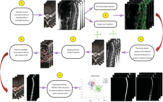
Method development.
Objectives
To develop a reliable protocol for automatic segmentation of Thoracolumbar spinal cord using MRI based on K-means clustering algorithm in 3D images.
Setting
University-based laboratory, Tehran, Iran.
Methods
T2 structural volumes acquired from the spinal cord of 20 uninjured volunteers on a 3T MR scanner. We proposed an automatic method for spinal cord segmentation based on the K-means clustering algorithm in 3D images and compare our results with two available segmentation methods (PropSeg, DeepSeg) implemented in the Spinal Cord Toolbox. Dice and Hausdorff were used to compare the results of our method (K-Seg) with the manual segmentation, PropSeg, and DeepSeg.
Results
The accuracy of our automatic segmentation method for T2-weighted images was significantly better or similar to the SCT methods, in terms of 3D DC (p < 0.001). The 3D DCs were respectively (0.81 ± 0.04) and Hausdorff Distance (12.3 ± 2.48) by the K-Seg method in contrary to other SCT methods for T2-weighted images.
Conclusions
The output with similar protocols showed that K-Seg results match the manual segmentation better than the other methods especially on the thoracolumbar levels in the spinal cord due to the low image contrast as a result of poor SNR in these areas.
中文翻译:
使用K均值聚类算法从T2加权MRI对胸腰椎脊髓和椎管进行全自动3D分割。
学习规划
方法开发。
目标
利用3D图像中基于K-means聚类算法的MRI,开发可靠的胸腰椎脊髓自动分割协议。
设置
基于大学的实验室,伊朗德黑兰。
方法
在3T MR扫描仪上从20名未受伤志愿者的脊髓获得的T2结构体积。我们提出了一种基于K-means聚类算法的3D图像自动脊髓分割方法,并将结果与脊髓工具箱中实现的两种可用分割方法(PropSeg,DeepSeg)进行了比较。使用Dice和Hausdorff将我们的方法(K-Seg)与手动分割,PropSeg和DeepSeg的结果进行比较。
结果
就3D DC而言,我们针对T2加权图像的自动分割方法的准确性明显优于或类似于SCT方法(p <0.001)。与其他T2加权图像的SCT方法相比,通过K-Seg方法的3D DC分别为(0.81±0.04)和Hausdorff距离(12.3±2.48)。
结论
具有类似协议的输出结果表明,由于这些区域的SNR较差,图像对比度低,因此K-Seg的结果比其他方法更好地匹配了手动分割,尤其是在脊髓的胸腰椎水平上。


 京公网安备 11010802027423号