当前位置:
X-MOL 学术
›
Compos. Sci. Technol.
›
论文详情
Our official English website, www.x-mol.net, welcomes your feedback! (Note: you will need to create a separate account there.)
Stereocomplex poly(lactic acid)-based composite nanofiber membranes with highly dispersed hydroxyapatite for potential bone tissue engineering
Composites Science and Technology ( IF 9.1 ) Pub Date : 2020-05-01 , DOI: 10.1016/j.compscitech.2020.108107 Di Chuan , Rangrang Fan , Yuelong Wang , Yangmei Ren , Chao Wang , Ying Du , Liangxue Zhou , Jie Yu , Yingchun Gu , Haifeng Chen , Gang Guo
Composites Science and Technology ( IF 9.1 ) Pub Date : 2020-05-01 , DOI: 10.1016/j.compscitech.2020.108107 Di Chuan , Rangrang Fan , Yuelong Wang , Yangmei Ren , Chao Wang , Ying Du , Liangxue Zhou , Jie Yu , Yingchun Gu , Haifeng Chen , Gang Guo
|
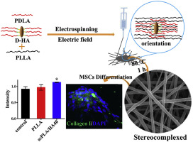
Abstract The development of materials with excellent mechanical properties and stem cell affinity for bone repair is a topic that has received significant interest in the field of bone tissue engineering. In this study, stereocomplex poly(lactic acid)-based composite nanofiber membranes that are compatible with bone marrow stem cells (BMSCs) were prepared with poly( d -lactic acid)-grafted hydroxyapatite and enantiomeric poly(lactic acid)s. The mechanical property tests showed that, compared with those from a heated poly( l -lactic acid) (PLLA) nanofiber membrane, the tensile strength and Young's modulus of the most strengthened composite nanofiber membrane herein increased by 30.16% and 34.56%, respectively. The X-ray diffraction results indicated that the stereocomplex crystallite (SC) of poly(lactic acid) formed in heated composite nanofiber membranes. The differential scanning calorimetry results showed that the melting temperature of the SC of heated composite nanofiber membranes was approximately 221 °C. Live/dead staining and scanning electron microscopy were used to observe the proliferation and adhesion of BMSCs cultured on the nanofiber membranes. Compared with the PLLA nanofiber membrane, increased type I collagen expression and improved formation of bone-like nodules can be observed after induction for nine days on the composite nanofiber membrane, indicating the potential application of the composite nanofiber membrane for bone tissue engineering.
中文翻译:

具有高度分散的羟基磷灰石的立体复合聚乳酸基复合纳米纤维膜用于潜在的骨组织工程
摘要 开发具有优异机械性能和干细胞亲和力的骨修复材料是骨组织工程领域的一个重要课题。在这项研究中,使用聚(d-乳酸)接枝的羟基磷灰石和对映体聚(乳酸)制备了与骨髓干细胞(BMSCs)相容的立体复合聚(乳酸)基复合纳米纤维膜。力学性能测试表明,与加热后的聚(l-乳酸)(PLLA)纳米纤维膜相比,本文中强度最高的复合纳米纤维膜的拉伸强度和杨氏模量分别提高了30.16%和34.56%。X 射线衍射结果表明聚乳酸的立体复合微晶 (SC) 在加热的复合纳米纤维膜中形成。差示扫描量热法结果表明,加热的复合纳米纤维膜的 SC 的熔化温度约为 221 °C。采用活/死染色和扫描电镜观察纳米纤维膜上培养的骨髓间充质干细胞的增殖和粘附情况。与 PLLA 纳米纤维膜相比,在复合纳米纤维膜上诱导 9 天后可以观察到 I 型胶原表达增加和骨样结节形成改善,表明复合纳米纤维膜在骨组织工程中的潜在应用。差示扫描量热法结果表明,加热后的复合纳米纤维膜的 SC 的熔化温度约为 221 °C。采用活/死染色和扫描电镜观察纳米纤维膜上培养的骨髓间充质干细胞的增殖和粘附情况。与 PLLA 纳米纤维膜相比,在复合纳米纤维膜上诱导 9 天后可以观察到 I 型胶原表达增加和骨样结节形成改善,表明复合纳米纤维膜在骨组织工程中的潜在应用。差示扫描量热法结果表明,加热后的复合纳米纤维膜的 SC 的熔化温度约为 221 °C。采用活/死染色和扫描电镜观察纳米纤维膜上培养的骨髓间充质干细胞的增殖和粘附情况。与 PLLA 纳米纤维膜相比,在复合纳米纤维膜上诱导 9 天后可以观察到 I 型胶原表达增加和骨样结节形成改善,表明复合纳米纤维膜在骨组织工程中的潜在应用。
更新日期:2020-05-01
中文翻译:
具有高度分散的羟基磷灰石的立体复合聚乳酸基复合纳米纤维膜用于潜在的骨组织工程
摘要 开发具有优异机械性能和干细胞亲和力的骨修复材料是骨组织工程领域的一个重要课题。在这项研究中,使用聚(d-乳酸)接枝的羟基磷灰石和对映体聚(乳酸)制备了与骨髓干细胞(BMSCs)相容的立体复合聚(乳酸)基复合纳米纤维膜。力学性能测试表明,与加热后的聚(l-乳酸)(PLLA)纳米纤维膜相比,本文中强度最高的复合纳米纤维膜的拉伸强度和杨氏模量分别提高了30.16%和34.56%。X 射线衍射结果表明聚乳酸的立体复合微晶 (SC) 在加热的复合纳米纤维膜中形成。差示扫描量热法结果表明,加热的复合纳米纤维膜的 SC 的熔化温度约为 221 °C。采用活/死染色和扫描电镜观察纳米纤维膜上培养的骨髓间充质干细胞的增殖和粘附情况。与 PLLA 纳米纤维膜相比,在复合纳米纤维膜上诱导 9 天后可以观察到 I 型胶原表达增加和骨样结节形成改善,表明复合纳米纤维膜在骨组织工程中的潜在应用。差示扫描量热法结果表明,加热后的复合纳米纤维膜的 SC 的熔化温度约为 221 °C。采用活/死染色和扫描电镜观察纳米纤维膜上培养的骨髓间充质干细胞的增殖和粘附情况。与 PLLA 纳米纤维膜相比,在复合纳米纤维膜上诱导 9 天后可以观察到 I 型胶原表达增加和骨样结节形成改善,表明复合纳米纤维膜在骨组织工程中的潜在应用。差示扫描量热法结果表明,加热后的复合纳米纤维膜的 SC 的熔化温度约为 221 °C。采用活/死染色和扫描电镜观察纳米纤维膜上培养的骨髓间充质干细胞的增殖和粘附情况。与 PLLA 纳米纤维膜相比,在复合纳米纤维膜上诱导 9 天后可以观察到 I 型胶原表达增加和骨样结节形成改善,表明复合纳米纤维膜在骨组织工程中的潜在应用。


 京公网安备 11010802027423号