当前位置:
X-MOL 学术
›
Acta Biomater.
›
论文详情
Our official English website, www.x-mol.net, welcomes your feedback! (Note: you will need to create a separate account there.)
Quantitative 3D structural analysis of the cellular microstructure of sea urchin spines (I): Methodology.
Acta Biomaterialia ( IF 9.7 ) Pub Date : 2020-02-25 , DOI: 10.1016/j.actbio.2020.02.034 Ting Yang 1 , Ziling Wu 2 , Hongshun Chen 1 , Yunhui Zhu 2 , Ling Li 1
Acta Biomaterialia ( IF 9.7 ) Pub Date : 2020-02-25 , DOI: 10.1016/j.actbio.2020.02.034 Ting Yang 1 , Ziling Wu 2 , Hongshun Chen 1 , Yunhui Zhu 2 , Ling Li 1
Affiliation
|
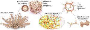
The mineralized skeletons of echinoderms are characterized by their complex, open-cell porous microstructure (also known as stereom), which exhibits vast variations in pore sizes, branch morphology, and three-dimensional (3D) organization patterns among different species. Quantitative description and analysis of these cellular structures in 3D are needed in order to understand their mechanical properties and underlying design strategies. In this paper series, we present a framework for analyzing such structures based on high-resolution 3D tomography data and utilize this framework to investigate the structural designs of stereom by using the spines from the sea urchin Heterocentrotus mamillatus as a model system. The first paper here reports the proposed cellular network analysis framework, which consists of five major steps: synchrotron-based tomography and hierarchical convolutional neural network-based reconstruction, machine learning-based segmentation, cellular network registration, feature extraction, and data representation and analysis. This framework enables the characterization of the porous stereom structures at the individual node and branch level (~10 µm), the local cellular level (~100 µm), and the global network level (~1 mm). We define and quantify multiple structural descriptors at each level, such as node connectivity, branch length and orientation, branch profile, ring structure, etc., which allows us to investigate the cellular network construction of H. mamillatus spines quantitatively. The methodology reported here could be tailored to analyze other natural or engineering open-cell porous materials for a comprehensive multiscale network representation and mechanical analysis. STATEMENT OF SIGNIFICANCE: The mechanical robustness of the biomineralized porous structures in sea urchin spines has long been recognized. However, quantitative cellular network representation and analysis of this class of natural cellular solids are still limited in the literature. This constrains our capability to fully understand the mechanical properties and design strategies in sea urchin spines and other similar echinoderms' porous skeletal structures. Combining high-resolution tomography and computer vision-based analysis, this work presents a multiscale 3D network analysis framework, which allows for extraction, registration, and quantification of sea urchin spines' complex porous structure from the individual branch and node level to the global network level. This 3D structural analysis is relevant to a diversity of research fields, such as biomineralization, skeletal biology, biomimetics, material science, etc.
中文翻译:

海胆刺细胞微结构的定量3D结构分析(I):方法论。
棘皮动物的矿化骨骼的特征是其复杂的开孔多孔微结构(也称为立体结构),在不同物种之间,其孔径,分支形态和三维(3D)组织模式表现出巨大差异。为了了解它们的机械性能和基本的设计策略,需要对3D中的这些细胞结构进行定量描述和分析。在本论文系列中,我们提供了一个基于高分辨率3D层析成像数据分析此类结构的框架,并利用该框架以海胆Heterocentrotus mamillatus的刺为模型系统来研究立体构造。这里的第一篇论文报告了建议的蜂窝网络分析框架,该框架包括五个主要步骤:基于同步加速器的层析成像和基于分层卷积神经网络的重建,基于机器学习的分割,蜂窝网络注册,特征提取以及数据表示和分析。这种框架可以表征单个节点和分支水平(〜10 µm),局部细胞水平(〜100 µm)和全局网络水平(〜1 mm)的多孔立体结构。我们在每个级别上定义和量化多个结构描述符,例如节点连接性,分支长度和方向,分支轮廓,环结构等,这使我们能够定量地研究H. mamillatus刺的细胞网络构造。此处报告的方法可以定制为分析其他天然或工程开孔多孔材料,以进行全面的多尺度网络表示和机械分析。意义声明:人们早已认识到海胆棘突中生物矿化多孔结构的机械强度。但是,在这类文献中,对这类天然细胞固体的定量细胞网络表示和分析仍然受到限制。这限制了我们充分了解海胆刺和其他类似棘皮动物的多孔骨骼结构的机械性能和设计策略的能力。结合高分辨率层析成像技术和基于计算机视觉的分析,这项工作提出了一种多尺度3D网络分析框架,该框架可进行提取,注册,并从单个分支和节点级别到全球网络级别量化海胆棘刺的复杂多孔结构。这种3D结构分析与多种研究领域相关,例如生物矿化,骨骼生物学,仿生生物,材料科学等。
更新日期:2020-02-25
中文翻译:
海胆刺细胞微结构的定量3D结构分析(I):方法论。
棘皮动物的矿化骨骼的特征是其复杂的开孔多孔微结构(也称为立体结构),在不同物种之间,其孔径,分支形态和三维(3D)组织模式表现出巨大差异。为了了解它们的机械性能和基本的设计策略,需要对3D中的这些细胞结构进行定量描述和分析。在本论文系列中,我们提供了一个基于高分辨率3D层析成像数据分析此类结构的框架,并利用该框架以海胆Heterocentrotus mamillatus的刺为模型系统来研究立体构造。这里的第一篇论文报告了建议的蜂窝网络分析框架,该框架包括五个主要步骤:基于同步加速器的层析成像和基于分层卷积神经网络的重建,基于机器学习的分割,蜂窝网络注册,特征提取以及数据表示和分析。这种框架可以表征单个节点和分支水平(〜10 µm),局部细胞水平(〜100 µm)和全局网络水平(〜1 mm)的多孔立体结构。我们在每个级别上定义和量化多个结构描述符,例如节点连接性,分支长度和方向,分支轮廓,环结构等,这使我们能够定量地研究H. mamillatus刺的细胞网络构造。此处报告的方法可以定制为分析其他天然或工程开孔多孔材料,以进行全面的多尺度网络表示和机械分析。意义声明:人们早已认识到海胆棘突中生物矿化多孔结构的机械强度。但是,在这类文献中,对这类天然细胞固体的定量细胞网络表示和分析仍然受到限制。这限制了我们充分了解海胆刺和其他类似棘皮动物的多孔骨骼结构的机械性能和设计策略的能力。结合高分辨率层析成像技术和基于计算机视觉的分析,这项工作提出了一种多尺度3D网络分析框架,该框架可进行提取,注册,并从单个分支和节点级别到全球网络级别量化海胆棘刺的复杂多孔结构。这种3D结构分析与多种研究领域相关,例如生物矿化,骨骼生物学,仿生生物,材料科学等。


 京公网安备 11010802027423号