当前位置:
X-MOL 学术
›
Adv. Healthcare Mater.
›
论文详情
Our official English website, www.x-mol.net, welcomes your feedback! (Note: you will need to create a separate account there.)
Recapitulating and Deciphering Tumor Microenvironment by Using 3D Printed Plastic Brick-Like Microfluidic Cell Patterning.
Advanced Healthcare Materials ( IF 10.0 ) Pub Date : 2020-02-24 , DOI: 10.1002/adhm.201901713 Yang Liu 1 , Yingying Liu 1 , Xiaonan Zheng 1 , Liang Zhao 1 , Xueji Zhang 1
Advanced Healthcare Materials ( IF 10.0 ) Pub Date : 2020-02-24 , DOI: 10.1002/adhm.201901713 Yang Liu 1 , Yingying Liu 1 , Xiaonan Zheng 1 , Liang Zhao 1 , Xueji Zhang 1
Affiliation
|
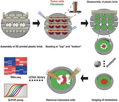
Within the body, tumor cells are surrounded by neighboring counterparts, such as extracellular matrix, vasculature, and host stroma, which is also known as the tumor microenvironment. To understand tumorigenesis, it is essential to reconstitute the incorporative tumor niche with quantitative measurements in vitro. Here, a 3D printed plastic brick-like microfluidic gadget is developed for spatially patterning tumors and fibroblasts, enabling the recapitulation of tumor microenvironment with minimized microfluidic expertise and compatibility of standard pipetting. This method facilitates heterotypic coculturing, quantitative phenotype decoding, and downstream molecular assays with a small number of cells (less than 100). Phenotypic and gene/protein expression-based analysis of cell-cell interactions between fibrosarcoma cells and fibroblasts on this device reveals that the tumor and its counterparts show reciprocal synergism mainly by upregulation of proinflammatory cytokines. Notably, at the whole transcriptional landscape (RNA-seq), fibroblasts display a transition from normal to cancer-associated fibroblast (CAF)-like phase, and tumor cells exhibit a hyperactive ribosome biogenesis. The mouse xenograft model is also involved to validate the in vitro analysis. Given its easy-to-use feature, full compatibility with molecular analysis, and open-source accessibility, this approach provides an in vitro experimental system to advance knowledge of tumorigenesis and the corresponding tumor microenvironment.
中文翻译:

通过使用3D打印的塑料砖样微流控细胞图案来概述和破译肿瘤微环境。
在体内,肿瘤细胞被周围的对应物包围,例如细胞外基质,脉管系统和宿主基质,这也被称为肿瘤微环境。要了解肿瘤发生,必须通过体外定量测量来重建合并的肿瘤位。在这里,开发了一种3D打印的塑料砖状微流控小工具,用于在空间上对肿瘤和成纤维细胞进行图案化,从而能够以最小化的微流控技术和标准移液的兼容性重现肿瘤微环境。这种方法有助于异型共培养,定量表型解码和少量细胞(少于100个)的下游分子测定。基于纤维细胞肉瘤细胞与成纤维细胞之间的细胞间相互作用的基于表型和基因/蛋白质表达的分析表明,肿瘤及其对应物主要通过促炎性细胞因子的上调表现出相互的协同作用。值得注意的是,在整个转录环境(RNA-seq)上,成纤维细胞显示出从正常到癌相关成纤维细胞(CAF)样阶段的转变,并且肿瘤细胞表现出过度活跃的核糖体生物发生。还涉及小鼠异种移植模型以验证体外分析。鉴于其易于使用的功能,与分子分析的完全兼容性以及开放源代码的可访问性,该方法提供了一种体外实验系统,以提高对肿瘤发生和相应的肿瘤微环境的认识。
更新日期:2020-03-19
中文翻译:
通过使用3D打印的塑料砖样微流控细胞图案来概述和破译肿瘤微环境。
在体内,肿瘤细胞被周围的对应物包围,例如细胞外基质,脉管系统和宿主基质,这也被称为肿瘤微环境。要了解肿瘤发生,必须通过体外定量测量来重建合并的肿瘤位。在这里,开发了一种3D打印的塑料砖状微流控小工具,用于在空间上对肿瘤和成纤维细胞进行图案化,从而能够以最小化的微流控技术和标准移液的兼容性重现肿瘤微环境。这种方法有助于异型共培养,定量表型解码和少量细胞(少于100个)的下游分子测定。基于纤维细胞肉瘤细胞与成纤维细胞之间的细胞间相互作用的基于表型和基因/蛋白质表达的分析表明,肿瘤及其对应物主要通过促炎性细胞因子的上调表现出相互的协同作用。值得注意的是,在整个转录环境(RNA-seq)上,成纤维细胞显示出从正常到癌相关成纤维细胞(CAF)样阶段的转变,并且肿瘤细胞表现出过度活跃的核糖体生物发生。还涉及小鼠异种移植模型以验证体外分析。鉴于其易于使用的功能,与分子分析的完全兼容性以及开放源代码的可访问性,该方法提供了一种体外实验系统,以提高对肿瘤发生和相应的肿瘤微环境的认识。


 京公网安备 11010802027423号