当前位置:
X-MOL 学术
›
ACS Chem. Neurosci.
›
论文详情
Our official English website, www.x-mol.net, welcomes your feedback! (Note: you will need to create a separate account there.)
Regional Variation in Striatal Dopamine Spillover and Release Plasticity.
ACS Chemical Neuroscience ( IF 5 ) Pub Date : 2020-02-19 , DOI: 10.1021/acschemneuro.9b00577 Seth H. Walters , Zhan Shu , Adrian C. Michael , Edwin S. Levitan
ACS Chemical Neuroscience ( IF 5 ) Pub Date : 2020-02-19 , DOI: 10.1021/acschemneuro.9b00577 Seth H. Walters , Zhan Shu , Adrian C. Michael , Edwin S. Levitan
|
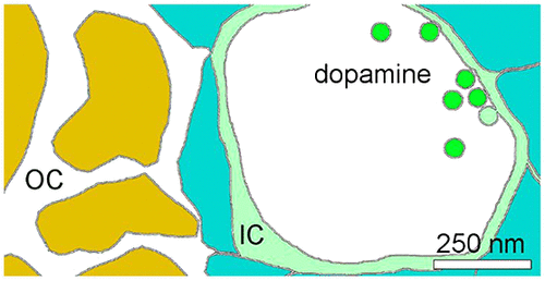
Recent optical observations of dopamine at axon terminals and kinetic modeling of evoked dopamine responses measured by fast scan cyclic voltammetry (FSCV) support local restriction of dopamine diffusion at synaptic release sites. Yet, how this diffusion barrier affects synaptic and volume transmission is unknown. Here, a deficiency in a previous kinetic model's fitting of stimulus trains is remedied by replacing an earlier assumption that dopamine transporters (DATs) are present only on the outer side of the diffusion barrier with the assumption that they are present on both sides. This is consistent with the known distribution of DATs, which does not show obvious DAT-free zones proximal to dopamine release sites. A simultaneous multifitting strategy is then shown to enable unique model fits to sets of evoked dopamine FSCV responses acquired in vivo or in brain slices. This data analysis technique permits, for the first time, the calculation of the fraction of dopamine which spills over from what appears to be the perisynaptic space, as well as other parameters such as dopamine release, release plasticity, and uptake. This analysis shows that dopamine's diffusion away from its release sites is remarkably hindered (τ = 5 s), but dopamine responses are rapid because of DAT activity. Furthermore, the new analysis reveals that uptake inhibitors can inhibit dopamine release during a stimulus train, apparently by depleting the releasable pool. It is suggested that ongoing uptake is critical for maintaining ongoing synaptic dopamine release and that the previously reported and also herein claimed increase of the initial dopamine release of some uptake inhibitors might be an important mechanism in addiction. Finally, brain mapping data reveal that the diffusion barrier is conserved, but there are variations in perisynaptic uptake, volume transmission, and release plasticity within the rat striatum. Therefore, an analysis paradigm is developed to quantify previously unmeasured features of brain dopaminergic transmission and to reveal regional functional differences among dopamine synapses.
中文翻译:

纹状体多巴胺溢出和释放可塑性的区域差异。
最近对轴突末端多巴胺的光学观察和通过快速扫描循环伏安法 (FSCV) 测量的诱发多巴胺反应的动力学模型支持突触释放位点多巴胺扩散的局部限制。然而,这种扩散屏障如何影响突触和体积传递尚不清楚。在这里,通过将多巴胺转运蛋白 (DAT) 仅存在于扩散屏障外侧的早期假设替换为它们存在于两侧的假设,可以弥补先前动力学模型对刺激列车的拟合不足。这与 DAT 的已知分布一致,DAT 在多巴胺释放位点附近没有显示明显的无 DAT 区域。然后显示同步多重拟合策略,使独特的模型适合在体内或脑切片中获得的诱发多巴胺 FSCV 反应集。这种数据分析技术首次允许计算从似乎是突触周围空间溢出的多巴胺分数,以及其他参数,如多巴胺释放、释放可塑性和摄取。该分析表明,多巴胺从其释放位点的扩散受到显着阻碍 (τ = 5 s),但由于 DAT 活动,多巴胺反应很快。此外,新的分析表明,摄取抑制剂可以抑制多巴胺在刺激过程中的释放,显然是通过耗尽可释放池来实现的。这表明持续摄取对于维持持续的突触多巴胺释放至关重要,并且先前报道和本文还声称增加一些摄取抑制剂的初始多巴胺释放可能是成瘾的重要机制。最后,大脑映射数据显示扩散屏障是守恒的,但大鼠纹状体内的突触周围摄取、体积传递和释放可塑性存在变化。因此,开发了一种分析范式来量化大脑多巴胺能传递以前未测量的特征,并揭示多巴胺突触之间的区域功能差异。大脑映射数据显示扩散屏障是守恒的,但大鼠纹状体内的突触周围摄取、体积传递和释放可塑性存在变化。因此,开发了一种分析范式来量化大脑多巴胺能传递以前未测量的特征,并揭示多巴胺突触之间的区域功能差异。大脑映射数据显示扩散屏障是守恒的,但大鼠纹状体内的突触周围摄取、体积传递和释放可塑性存在变化。因此,开发了一种分析范式来量化大脑多巴胺能传递以前未测量的特征,并揭示多巴胺突触之间的区域功能差异。
更新日期:2020-02-28
中文翻译:
纹状体多巴胺溢出和释放可塑性的区域差异。
最近对轴突末端多巴胺的光学观察和通过快速扫描循环伏安法 (FSCV) 测量的诱发多巴胺反应的动力学模型支持突触释放位点多巴胺扩散的局部限制。然而,这种扩散屏障如何影响突触和体积传递尚不清楚。在这里,通过将多巴胺转运蛋白 (DAT) 仅存在于扩散屏障外侧的早期假设替换为它们存在于两侧的假设,可以弥补先前动力学模型对刺激列车的拟合不足。这与 DAT 的已知分布一致,DAT 在多巴胺释放位点附近没有显示明显的无 DAT 区域。然后显示同步多重拟合策略,使独特的模型适合在体内或脑切片中获得的诱发多巴胺 FSCV 反应集。这种数据分析技术首次允许计算从似乎是突触周围空间溢出的多巴胺分数,以及其他参数,如多巴胺释放、释放可塑性和摄取。该分析表明,多巴胺从其释放位点的扩散受到显着阻碍 (τ = 5 s),但由于 DAT 活动,多巴胺反应很快。此外,新的分析表明,摄取抑制剂可以抑制多巴胺在刺激过程中的释放,显然是通过耗尽可释放池来实现的。这表明持续摄取对于维持持续的突触多巴胺释放至关重要,并且先前报道和本文还声称增加一些摄取抑制剂的初始多巴胺释放可能是成瘾的重要机制。最后,大脑映射数据显示扩散屏障是守恒的,但大鼠纹状体内的突触周围摄取、体积传递和释放可塑性存在变化。因此,开发了一种分析范式来量化大脑多巴胺能传递以前未测量的特征,并揭示多巴胺突触之间的区域功能差异。大脑映射数据显示扩散屏障是守恒的,但大鼠纹状体内的突触周围摄取、体积传递和释放可塑性存在变化。因此,开发了一种分析范式来量化大脑多巴胺能传递以前未测量的特征,并揭示多巴胺突触之间的区域功能差异。大脑映射数据显示扩散屏障是守恒的,但大鼠纹状体内的突触周围摄取、体积传递和释放可塑性存在变化。因此,开发了一种分析范式来量化大脑多巴胺能传递以前未测量的特征,并揭示多巴胺突触之间的区域功能差异。


 京公网安备 11010802027423号