Nature Immunology ( IF 30.5 ) Pub Date : 2020-02-17 , DOI: 10.1038/s41590-020-0596-6 Marco De Giovanni 1, 2 , Valeria Cutillo 1, 2 , Amir Giladi 3 , Eleonora Sala 1, 2 , Carmela G Maganuco 1 , Chiara Medaglia 3 , Pietro Di Lucia 1 , Elisa Bono 1 , Claudia Cristofani 1 , Eleonora Consolo 1, 2 , Leonardo Giustini 1 , Alessandra Fiore 2 , Sarah Eickhoff 4 , Wolfgang Kastenmüller 4 , Ido Amit 3 , Mirela Kuka 1, 2 , Matteo Iannacone 1, 2, 5
|
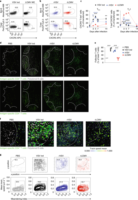
Differentiation of CD4+ T cells into either follicular helper T (TFH) or type 1 helper T (TH1) cells influences the balance between humoral and cellular adaptive immunity, but the mechanisms whereby pathogens elicit distinct effector cells are incompletely understood. Here we analyzed the spatiotemporal dynamics of CD4+ T cells during infection with recombinant vesicular stomatitis virus (VSV), which induces early, potent neutralizing antibodies, or recombinant lymphocytic choriomeningitis virus (LCMV), which induces a vigorous cellular response but inefficient neutralizing antibodies, expressing the same T cell epitope. Early exposure of dendritic cells to type I interferon (IFN), which occurred during infection with VSV, induced production of the cytokine IL-6 and drove TFH cell polarization, whereas late exposure to type I IFN, which occurred during infection with LCMV, did not induce IL-6 and allowed differentiation into TH1 cells. Thus, tight spatiotemporal regulation of type I IFN shapes antiviral CD4+ T cell differentiation and might instruct vaccine design strategies.
中文翻译:
I型干扰素表达的时空调控决定了CD4+ T细胞的抗病毒极化
CD4 + T 细胞分化为滤泡辅助 T (T FH ) 或 1 型辅助 T (T H 1) 细胞会影响体液和细胞适应性免疫之间的平衡,但病原体引发不同效应细胞的机制尚不完全清楚。这里我们分析了CD4 +的时空动态感染重组水泡性口炎病毒 (VSV) 期间的 T 细胞,可诱导早期有效的中和抗体,或重组淋巴细胞脉络丛脑膜炎病毒 (LCMV),可诱导强烈的细胞反应但中和抗体无效,表达相同的 T 细胞表位。树突状细胞早期暴露于 I 型干扰素 (IFN),发生在 VSV 感染期间,诱导细胞因子 IL-6 的产生并驱动 T FH细胞极化,而晚期暴露于 I 型干扰素,发生在 LCMV 感染期间,不诱导 IL-6 并允许分化为 T H 1 细胞。因此,I 型 IFN 的严格时空调控形成抗病毒 CD4 +T 细胞分化并可能指导疫苗设计策略。


 京公网安备 11010802027423号