当前位置:
X-MOL 学术
›
Adv. Mater. Technol.
›
论文详情
Our official English website, www.x-mol.net, welcomes your feedback! (Note: you will need to create a separate account there.)
Probing the Ultrastructure of Spheroids and Their Uptake of Magnetic Nanoparticles by FIB–SEM
Advanced Materials Technologies ( IF 6.8 ) Pub Date : 2020-02-17 , DOI: 10.1002/admt.201900687 Valentina Mollo 1 , Paola Scognamiglio 1 , Attilio Marino 2 , Gianni Ciofani 2, 3 , Francesca Santoro 1
Advanced Materials Technologies ( IF 6.8 ) Pub Date : 2020-02-17 , DOI: 10.1002/admt.201900687 Valentina Mollo 1 , Paola Scognamiglio 1 , Attilio Marino 2 , Gianni Ciofani 2, 3 , Francesca Santoro 1
Affiliation
|
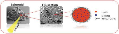
Spheroids are 3D cellular systems largely adopted as model for high‐throughput screening of molecules and diagnostics tools. Furthermore, those cellular platforms also represent a model for testing new delivery carries for selective targeting. The coupling between the 3D cell environment and the nanovectors can be explored at the macroscale by optical microscopy. However, the nanomaterial‐cell interplay finds major action at the single cell and extracellular matrix level with nanoscale interactions. Electron microscopy offers the resolution to investigate those interactions; however, the specimen preparation finds major drawbacks in its operation time and preciseness. In this context, focused ion beam and scanning electron microscopy (FIB–SEM) allows for fast processing and high resolution of the cell‐nanomaterial interface. Here, in fact, a novel approach is shown to prepare large‐area 3D spheroid cell culture specimens for FIB–SEM. Sectioning procedures are explored to preserve the peculiar structure of spheroids and their interaction with magnetic nanovectors. The results pave the way for advanced investigations of 3D cellular systems with nano and micromaterials relevant to tissue engineering, bioelectronics, and diagnostics.
中文翻译:

通过FIB–SEM探测球体的超微结构及其对磁性纳米颗粒的吸收
球体是3D细胞系统,被广泛用作分子高通量筛选和诊断工具的模型。此外,那些蜂窝平台还代表了一种模型,用于测试用于选择性靶向的新递送载体。3D细胞环境与纳米载体之间的耦合可以通过光学显微镜在宏观上进行探索。然而,纳米材料与细胞的相互作用在纳米级相互作用的单细胞和细胞外基质水平上发现了主要作用。电子显微镜提供了研究这些相互作用的分辨率。但是,样品制备在操作时间和准确性上存在主要缺陷。在这种情况下,聚焦离子束和扫描电子显微镜(FIB–SEM)可以实现细胞-纳米材料界面的快速处理和高分辨率。实际上,在这里 显示了一种新颖的方法可以制备用于FIB–SEM的大面积3D球形细胞培养标本。探索切片程序以保留球体的特殊结构及其与磁性纳米载体的相互作用。该结果为与组织工程,生物电子学和诊断学相关的纳米和微材料的3D细胞系统的高级研究铺平了道路。
更新日期:2020-03-09
中文翻译:
通过FIB–SEM探测球体的超微结构及其对磁性纳米颗粒的吸收
球体是3D细胞系统,被广泛用作分子高通量筛选和诊断工具的模型。此外,那些蜂窝平台还代表了一种模型,用于测试用于选择性靶向的新递送载体。3D细胞环境与纳米载体之间的耦合可以通过光学显微镜在宏观上进行探索。然而,纳米材料与细胞的相互作用在纳米级相互作用的单细胞和细胞外基质水平上发现了主要作用。电子显微镜提供了研究这些相互作用的分辨率。但是,样品制备在操作时间和准确性上存在主要缺陷。在这种情况下,聚焦离子束和扫描电子显微镜(FIB–SEM)可以实现细胞-纳米材料界面的快速处理和高分辨率。实际上,在这里 显示了一种新颖的方法可以制备用于FIB–SEM的大面积3D球形细胞培养标本。探索切片程序以保留球体的特殊结构及其与磁性纳米载体的相互作用。该结果为与组织工程,生物电子学和诊断学相关的纳米和微材料的3D细胞系统的高级研究铺平了道路。


 京公网安备 11010802027423号