Nature Machine Intelligence ( IF 23.8 ) Pub Date : 2020-02-14 , DOI: 10.1038/s42256-020-0154-9 Qi Yan 1 , Daniel E Weeks 2, 3 , Hongyi Xin 1 , Anand Swaroop 4 , Emily Y Chew 5 , Heng Huang 6 , Ying Ding 3 , Wei Chen 1, 2, 3
|
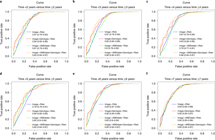
Both genetic and environmental factors influence the etiology of age-related macular degeneration (AMD), a leading cause of blindness. AMD severity is primarily measured by images of the fundus of the retina and recently developed machine learning methods can successfully predict AMD progression using image data. However, none of these methods have used both genetic and image data for predicting AMD progression. Here we used both genotypes and fundus images to predict whether an eye had progressed to late AMD with a modified deep convolutional neural network. In total, we used 31,262 fundus images and 52 AMD-associated genetic variants from 1,351 subjects from the Age-Related Eye Disease Study, which provided disease severity phenotypes and fundus images available at baseline and follow-up visits over a period of 12 years. Our results showed that fundus images coupled with genotypes could predict late AMD progression with an averaged area-under-the-curve value of 0.85 (95% confidence interval 0.83–0.86). The results using fundus images alone showed an averaged area under the receiver operating characteristic curve value of 0.81 (95% confidence interval 0.80–0.83). We implemented our model in a cloud-based application for individual risk assessment.
A preprint version of the article is available at ArXiv.中文翻译:
基于深度学习的与年龄相关的黄斑变性进展的预测。
遗传和环境因素都会影响与年龄有关的黄斑变性(AMD)的病因,而黄斑变性是致盲的主要原因。AMD严重性主要通过视网膜眼底图像来衡量,最近开发的机器学习方法可以使用图像数据成功预测AMD进程。但是,这些方法都没有使用遗传和图像数据来预测AMD进展。在这里,我们使用基因型和眼底图像来预测眼睛是否已经发展到具有改良的深度卷积神经网络的晚期AMD。我们总共使用了来自年龄相关性眼病研究的1,351名受试者的31,262个眼底图像和52个与AMD相关的遗传变异,这些研究提供了在12年内的基线和随访期间可获得的疾病严重程度表型和眼底图像。我们的结果表明,眼底图像与基因型结合可以预测晚期AMD进展,曲线下平均面积值为0.85(95%置信区间0.83-0.86)。仅使用眼底图像的结果显示,接收器工作特征曲线值下的平均面积为0.81(95%置信区间0.80-0.83)。我们在基于云的应用程序中实施了模型,用于个人风险评估。
该文章的预印本可从ArXiv获得。


 京公网安备 11010802027423号