当前位置:
X-MOL 学术
›
Nat. Protoc.
›
论文详情
Our official English website, www.x-mol.net, welcomes your feedback! (Note: you will need to create a separate account there.)
Superresolution imaging of chromatin fibers to visualize epigenetic information on replicative DNA.
Nature Protocols ( IF 14.8 ) Pub Date : 2020-02-12 , DOI: 10.1038/s41596-019-0283-y Matthew Wooten 1 , Yingying Li 1 , Jonathan Snedeker 1 , Zehra F Nizami 2 , Joseph G Gall 2 , Xin Chen 1
Nature Protocols ( IF 14.8 ) Pub Date : 2020-02-12 , DOI: 10.1038/s41596-019-0283-y Matthew Wooten 1 , Yingying Li 1 , Jonathan Snedeker 1 , Zehra F Nizami 2 , Joseph G Gall 2 , Xin Chen 1
Affiliation
|
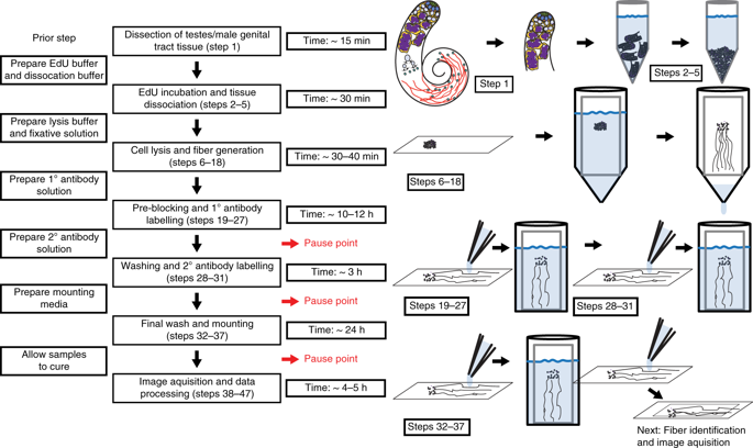
During DNA replication, the genetic information of a cell is copied. Subsequently, identical genetic information is segregated reliably to the two daughter cells through cell division. Meanwhile, DNA replication is intrinsically linked to the process of chromatin duplication, which is required for regulating gene expression and establishing cell identities. Understanding how chromatin is established, maintained or changed during DNA replication represents a fundamental question in biology. Recently, we developed a method to directly visualize chromatin components at individual replication forks undergoing DNA replication. This method builds upon the existing chromatin fiber technique and combines it with cell type-specific chromatin labeling and superresolution microscopy. In this method, a short pulse of nucleoside analog labels replicative regions in the cells of interest. Chromatin fibers are subsequently isolated and attached to a glass slide, after which a laminar flow of lysis buffer extends the lysed chromatin fibers parallel with the direction of the flow. Fibers are then immunostained for different chromatin-associated proteins and mounted for visualization using superresolution microscopy. Replication foci, or 'bubbles,' are identified by the presence of the incorporated nucleoside analog. For researchers experienced in molecular biology and superresolution microscopy, this protocol typically takes 2-3 d from sample preparation to data acquisition, with an additional day for data processing and quantification.
中文翻译:

染色质纤维的超分辨率成像以可视化复制 DNA 的表观遗传信息。
在 DNA 复制过程中,细胞的遗传信息被复制。随后,相同的遗传信息通过细胞分裂可靠地分离到两个子细胞。同时,DNA复制与染色质复制过程有着内在联系,染色质复制是调节基因表达和建立细胞身份所必需的。了解在 DNA 复制过程中染色质是如何建立、维持或改变的,是生物学中的一个基本问题。最近,我们开发了一种方法,可以直接可视化进行 DNA 复制的单个复制叉上的染色质成分。该方法建立在现有染色质纤维技术的基础上,并将其与细胞类型特异性染色质标记和超分辨率显微镜相结合。在这种方法中,核苷类似物的短脉冲标记感兴趣细胞中的复制区域。随后分离染色质纤维并将其连接到载玻片上,之后裂解缓冲液的层流将裂解的染色质纤维平行于流动方向延伸。然后针对不同的染色质相关蛋白对纤维进行免疫染色,并使用超分辨率显微镜进行可视化。复制焦点或“气泡”通过掺入的核苷类似物的存在来识别。对于在分子生物学和超分辨率显微镜方面经验丰富的研究人员,该协议通常需要 2-3 天从样品制备到数据采集,另外一天用于数据处理和量化。之后,裂解缓冲液的层流使裂解的染色质纤维平行于流动方向延伸。然后针对不同的染色质相关蛋白对纤维进行免疫染色,并使用超分辨率显微镜进行可视化。复制焦点或“气泡”通过掺入的核苷类似物的存在来识别。对于在分子生物学和超分辨率显微镜方面经验丰富的研究人员,该协议通常需要 2-3 天从样品制备到数据采集,另外一天用于数据处理和量化。之后,裂解缓冲液的层流使裂解的染色质纤维平行于流动方向延伸。然后针对不同的染色质相关蛋白对纤维进行免疫染色,并使用超分辨率显微镜进行可视化。复制焦点或“气泡”通过掺入的核苷类似物的存在来识别。对于在分子生物学和超分辨率显微镜方面经验丰富的研究人员,该协议通常需要 2-3 天从样品制备到数据采集,另外一天用于数据处理和量化。复制焦点或“气泡”通过掺入的核苷类似物的存在来识别。对于在分子生物学和超分辨率显微镜方面经验丰富的研究人员,该协议通常需要 2-3 天从样品制备到数据采集,另外一天用于数据处理和量化。复制焦点或“气泡”通过掺入的核苷类似物的存在来识别。对于在分子生物学和超分辨率显微镜方面经验丰富的研究人员,该协议通常需要 2-3 天从样品制备到数据采集,另外一天用于数据处理和量化。
更新日期:2020-02-12
中文翻译:
染色质纤维的超分辨率成像以可视化复制 DNA 的表观遗传信息。
在 DNA 复制过程中,细胞的遗传信息被复制。随后,相同的遗传信息通过细胞分裂可靠地分离到两个子细胞。同时,DNA复制与染色质复制过程有着内在联系,染色质复制是调节基因表达和建立细胞身份所必需的。了解在 DNA 复制过程中染色质是如何建立、维持或改变的,是生物学中的一个基本问题。最近,我们开发了一种方法,可以直接可视化进行 DNA 复制的单个复制叉上的染色质成分。该方法建立在现有染色质纤维技术的基础上,并将其与细胞类型特异性染色质标记和超分辨率显微镜相结合。在这种方法中,核苷类似物的短脉冲标记感兴趣细胞中的复制区域。随后分离染色质纤维并将其连接到载玻片上,之后裂解缓冲液的层流将裂解的染色质纤维平行于流动方向延伸。然后针对不同的染色质相关蛋白对纤维进行免疫染色,并使用超分辨率显微镜进行可视化。复制焦点或“气泡”通过掺入的核苷类似物的存在来识别。对于在分子生物学和超分辨率显微镜方面经验丰富的研究人员,该协议通常需要 2-3 天从样品制备到数据采集,另外一天用于数据处理和量化。之后,裂解缓冲液的层流使裂解的染色质纤维平行于流动方向延伸。然后针对不同的染色质相关蛋白对纤维进行免疫染色,并使用超分辨率显微镜进行可视化。复制焦点或“气泡”通过掺入的核苷类似物的存在来识别。对于在分子生物学和超分辨率显微镜方面经验丰富的研究人员,该协议通常需要 2-3 天从样品制备到数据采集,另外一天用于数据处理和量化。之后,裂解缓冲液的层流使裂解的染色质纤维平行于流动方向延伸。然后针对不同的染色质相关蛋白对纤维进行免疫染色,并使用超分辨率显微镜进行可视化。复制焦点或“气泡”通过掺入的核苷类似物的存在来识别。对于在分子生物学和超分辨率显微镜方面经验丰富的研究人员,该协议通常需要 2-3 天从样品制备到数据采集,另外一天用于数据处理和量化。复制焦点或“气泡”通过掺入的核苷类似物的存在来识别。对于在分子生物学和超分辨率显微镜方面经验丰富的研究人员,该协议通常需要 2-3 天从样品制备到数据采集,另外一天用于数据处理和量化。复制焦点或“气泡”通过掺入的核苷类似物的存在来识别。对于在分子生物学和超分辨率显微镜方面经验丰富的研究人员,该协议通常需要 2-3 天从样品制备到数据采集,另外一天用于数据处理和量化。


 京公网安备 11010802027423号