Our official English website, www.x-mol.net, welcomes your feedback! (Note: you will need to create a separate account there.)
Absence of microglia or presence of peripherally-derived macrophages does not affect tau pathology in young or old hTau mice.
Glia ( IF 6.2 ) Pub Date : 2020-02-10 , DOI: 10.1002/glia.23794 Keying Zhu 1 , Melanie Pieber 1 , Jinming Han 1 , Klas Blomgren 2, 3 , Xing-Mei Zhang 1 , Robert A Harris 1 , Harald Lund 1, 4, 5
Glia ( IF 6.2 ) Pub Date : 2020-02-10 , DOI: 10.1002/glia.23794 Keying Zhu 1 , Melanie Pieber 1 , Jinming Han 1 , Klas Blomgren 2, 3 , Xing-Mei Zhang 1 , Robert A Harris 1 , Harald Lund 1, 4, 5
Affiliation
|
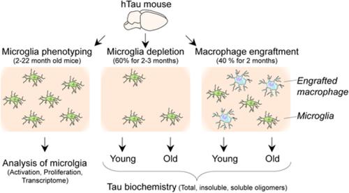
Microglia are implicated in the pathophysiology of several neurodegenerative disorders, including Alzheimer's disease. While the role of microglia and peripheral macrophages in regulating amyloid beta pathology has been well characterized, the impact of these distinct cell subsets on tau pathology remains poorly understood. We and others have recently demonstrated that monocytes can engraft the brain and give rise to long-lived parenchymal macrophages, even under nonpathological conditions. We undertook the current study to investigate the regulation of tau pathology by microglia and peripheral macrophages using hTau transgenic mice, which do not exhibit microglial activation/pathology or macrophage engraftment. To assess the direct impact of microglia on tau pathology we developed a protocol for long-term microglial depletion in Cx3cr1CreER R26DTA mice and crossed them with hTau mice. We then depleted microglia up to 3 months in both young and old mice, but no net change in forebrain soluble oligomeric tau or total or phosphorylated levels of aggregated tau was recorded. To investigate the consequence of peripherally-derived parenchymal macrophages on tau aggregation we partially repopulated the hTau microglial pool with peripheral macrophages, but this also did not affect levels of tau oligomers or insoluble aggregates. Our study questions the direct involvement of microglia or peripheral macrophages in the development of tau pathology in the hTau model.
中文翻译:

小胶质细胞的缺失或外周衍生的巨噬细胞的存在不会影响年轻或年老 hTau 小鼠的 tau 病理学。
小胶质细胞与多种神经退行性疾病的病理生理学有关,包括阿尔茨海默病。虽然小胶质细胞和外周巨噬细胞在调节淀粉样蛋白 β 病理学中的作用已经得到很好的表征,但这些不同的细胞亚群对 tau 病理学的影响仍然知之甚少。我们和其他人最近证明,即使在非病理条件下,单核细胞也可以植入大脑并产生长寿的实质巨噬细胞。我们进行了目前的研究,使用 hTau 转基因小鼠研究小胶质细胞和外周巨噬细胞对 tau 病理的调节,这些小鼠不表现出小胶质细胞激活/病理或巨噬细胞移植。为了评估小胶质细胞对 tau 病理学的直接影响,我们制定了 Cx3cr1CreER R26DTA 小鼠中长期小胶质细胞耗竭的方案,并将它们与 hTau 小鼠杂交。然后,我们在年轻和年老小鼠中耗尽小胶质细胞长达 3 个月,但没有记录前脑可溶性寡聚 tau 或聚合 tau 的总或磷酸化水平的净变化。为了研究外周衍生的实质巨噬细胞对 tau 聚集的影响,我们用外周巨噬细胞部分重新填充 hTau 小胶质细胞池,但这也不影响 tau 寡聚体或不溶性聚集体的水平。我们的研究质疑小胶质细胞或外周巨噬细胞在 hTau 模型中 tau 病理学的发展中的直接参与。然后,我们在年轻和年老小鼠中耗尽小胶质细胞长达 3 个月,但没有记录前脑可溶性寡聚 tau 或聚合 tau 的总或磷酸化水平的净变化。为了研究外周衍生的实质巨噬细胞对 tau 聚集的影响,我们用外周巨噬细胞部分重新填充 hTau 小胶质细胞池,但这也不影响 tau 寡聚体或不溶性聚集体的水平。我们的研究质疑小胶质细胞或外周巨噬细胞在 hTau 模型中 tau 病理学的发展中的直接参与。然后,我们在年轻和年老小鼠中耗尽小胶质细胞长达 3 个月,但没有记录前脑可溶性寡聚 tau 或聚合 tau 的总或磷酸化水平的净变化。为了研究外周衍生的实质巨噬细胞对 tau 聚集的影响,我们用外周巨噬细胞部分重新填充 hTau 小胶质细胞池,但这也不影响 tau 寡聚体或不溶性聚集体的水平。我们的研究质疑小胶质细胞或外周巨噬细胞在 hTau 模型中 tau 病理学的发展中的直接参与。为了研究外周衍生的实质巨噬细胞对 tau 聚集的影响,我们用外周巨噬细胞部分重新填充 hTau 小胶质细胞池,但这也不影响 tau 寡聚体或不溶性聚集体的水平。我们的研究质疑小胶质细胞或外周巨噬细胞在 hTau 模型中 tau 病理学的发展中的直接参与。为了研究外周衍生的实质巨噬细胞对 tau 聚集的影响,我们用外周巨噬细胞部分重新填充 hTau 小胶质细胞池,但这也不影响 tau 寡聚体或不溶性聚集体的水平。我们的研究质疑小胶质细胞或外周巨噬细胞在 hTau 模型中 tau 病理学的发展中的直接参与。
更新日期:2020-02-10
中文翻译:
小胶质细胞的缺失或外周衍生的巨噬细胞的存在不会影响年轻或年老 hTau 小鼠的 tau 病理学。
小胶质细胞与多种神经退行性疾病的病理生理学有关,包括阿尔茨海默病。虽然小胶质细胞和外周巨噬细胞在调节淀粉样蛋白 β 病理学中的作用已经得到很好的表征,但这些不同的细胞亚群对 tau 病理学的影响仍然知之甚少。我们和其他人最近证明,即使在非病理条件下,单核细胞也可以植入大脑并产生长寿的实质巨噬细胞。我们进行了目前的研究,使用 hTau 转基因小鼠研究小胶质细胞和外周巨噬细胞对 tau 病理的调节,这些小鼠不表现出小胶质细胞激活/病理或巨噬细胞移植。为了评估小胶质细胞对 tau 病理学的直接影响,我们制定了 Cx3cr1CreER R26DTA 小鼠中长期小胶质细胞耗竭的方案,并将它们与 hTau 小鼠杂交。然后,我们在年轻和年老小鼠中耗尽小胶质细胞长达 3 个月,但没有记录前脑可溶性寡聚 tau 或聚合 tau 的总或磷酸化水平的净变化。为了研究外周衍生的实质巨噬细胞对 tau 聚集的影响,我们用外周巨噬细胞部分重新填充 hTau 小胶质细胞池,但这也不影响 tau 寡聚体或不溶性聚集体的水平。我们的研究质疑小胶质细胞或外周巨噬细胞在 hTau 模型中 tau 病理学的发展中的直接参与。然后,我们在年轻和年老小鼠中耗尽小胶质细胞长达 3 个月,但没有记录前脑可溶性寡聚 tau 或聚合 tau 的总或磷酸化水平的净变化。为了研究外周衍生的实质巨噬细胞对 tau 聚集的影响,我们用外周巨噬细胞部分重新填充 hTau 小胶质细胞池,但这也不影响 tau 寡聚体或不溶性聚集体的水平。我们的研究质疑小胶质细胞或外周巨噬细胞在 hTau 模型中 tau 病理学的发展中的直接参与。然后,我们在年轻和年老小鼠中耗尽小胶质细胞长达 3 个月,但没有记录前脑可溶性寡聚 tau 或聚合 tau 的总或磷酸化水平的净变化。为了研究外周衍生的实质巨噬细胞对 tau 聚集的影响,我们用外周巨噬细胞部分重新填充 hTau 小胶质细胞池,但这也不影响 tau 寡聚体或不溶性聚集体的水平。我们的研究质疑小胶质细胞或外周巨噬细胞在 hTau 模型中 tau 病理学的发展中的直接参与。为了研究外周衍生的实质巨噬细胞对 tau 聚集的影响,我们用外周巨噬细胞部分重新填充 hTau 小胶质细胞池,但这也不影响 tau 寡聚体或不溶性聚集体的水平。我们的研究质疑小胶质细胞或外周巨噬细胞在 hTau 模型中 tau 病理学的发展中的直接参与。为了研究外周衍生的实质巨噬细胞对 tau 聚集的影响,我们用外周巨噬细胞部分重新填充 hTau 小胶质细胞池,但这也不影响 tau 寡聚体或不溶性聚集体的水平。我们的研究质疑小胶质细胞或外周巨噬细胞在 hTau 模型中 tau 病理学的发展中的直接参与。


 京公网安备 11010802027423号