Journal of Allergy and Clinical Immunology ( IF 14.2 ) Pub Date : 2020-02-07 , DOI: 10.1016/j.jaci.2020.01.042 Helen He 1 , Hemant Suryawanshi 2 , Pavel Morozov 2 , Jesús Gay-Mimbrera 3 , Ester Del Duca 1 , Hyun Je Kim 1 , Naoya Kameyama 1 , Yeriel Estrada 1 , Evan Der 4 , James G Krueger 5 , Juan Ruano 3 , Thomas Tuschl 2 , Emma Guttman-Yassky 1
|
Background
Atopic dermatitis (AD) is a prevalent inflammatory skin disease with a complex pathogenesis involving immune cell and epidermal abnormalities. Despite whole tissue biopsy studies that have advanced the mechanistic understanding of AD, single cell–based molecular alterations are largely unknown.
Objective
Our aims were to construct a detailed, high-resolution atlas of cell populations and assess variability in cell composition and cell-specific gene expression in the skin of patients with AD versus in controls.
Methods
We performed single-cell RNA sequencing on skin biopsy specimens from 5 patients with AD (4 lesional samples and 5 nonlesional samples) and 7 healthy control subjects, using 10× Genomics.
Results
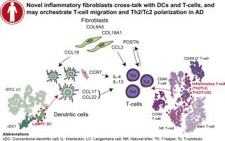
We created transcriptomic profiles for 39,042 AD (lesional and nonlesional) and healthy skin cells. Fibroblasts demonstrated a novel COL6A5+COL18A1+ subpopulation that was unique to lesional AD and expressed CCL2 and CCL19 cytokines. A corresponding LAMP3+ dendritic cell (DC) population that expressed the CCL19 receptor CCR7 was also unique to AD lesions, illustrating a potential role for fibroblast signaling to immune cells. The lesional AD samples were characterized by expansion of inflammatory DCs (CD1A+FCER1A+) and tissue-resident memory T cells (CD69+CD103+). The frequencies of type 2 (IL13+)/type 22 (IL22+) T cells were higher than those of type 1 (IFNG+) in lesional AD, whereas this ratio was slightly diminished in nonlesional AD and further diminished in controls.
Conclusion
AD lesions were characterized by expanded type 2/type 22 T cells and inflammatory DCs, and by a unique inflammatory fibroblast that may interact with immune cells to regulate lymphoid cell organization and type 2 inflammation.
中文翻译:
人皮肤的单细胞转录组分析确定了特应性皮炎中新的成纤维细胞亚群和免疫亚群的富集。
背景
特应性皮炎(AD)是一种常见的炎症性皮肤病,发病机制复杂,涉及免疫细胞和表皮异常。尽管整个组织活检研究已经提高了对AD的机制的了解,但基于单细胞的分子改变在很大程度上还是未知的。
目的
我们的目标是构建详细的高分辨率细胞群图集,并评估AD患者与对照组患者皮肤中细胞组成和细胞特异性基因表达的差异。
方法
我们使用10x基因组学对5名AD患者(4个病灶样品和5个非病灶样品)和7个健康对照受试者的皮肤活检标本进行了单细胞RNA测序。
结果
我们创建了39,042 AD(病变和非病变)和健康皮肤细胞的转录组谱。成纤维细胞显示出新颖的COL6A5 + COL18A1 +亚群,是病变AD特有的,并表达CCL2和CCL19细胞因子。表达CCL19受体CCR7的相应LAMP3 +树突状细胞(DC)群体也是AD病变所独有的,说明成纤维细胞信号传导至免疫细胞的潜在作用。皮损AD样品通过炎症的DC(扩张特征CD1A + FCER1A +)和组织驻留记忆T细胞(CD69+ CD103 +)。病变AD中2型(IL13 +)/ 22型(IL22 +)T细胞的频率高于1型(IFNG +),而在非病变AD中该比例略有降低,而在对照组中则进一步降低。
结论
AD病变的特征在于扩大的2/2型22 T细胞和炎性DC,以及独特的炎性成纤维细胞,其可能与免疫细胞相互作用以调节淋巴样细胞的组织和2型炎症。


 京公网安备 11010802027423号