当前位置:
X-MOL 学术
›
J. Biophotonics
›
论文详情
Our official English website, www.x-mol.net, welcomes your feedback! (Note: you will need to create a separate account there.)
In vivo detection of venous sinus distension due to intracranial hypotension in small animal using pulsed-laser-diode photoacoustic tomography.
Journal of Biophotonics ( IF 2.8 ) Pub Date : 2020-03-20 , DOI: 10.1002/jbio.201960162 Praveenbalaji Rajendran 1 , Samiran Sahu 1 , Rhonnie Austria Dienzo 1 , Manojit Pramanik 1
Journal of Biophotonics ( IF 2.8 ) Pub Date : 2020-03-20 , DOI: 10.1002/jbio.201960162 Praveenbalaji Rajendran 1 , Samiran Sahu 1 , Rhonnie Austria Dienzo 1 , Manojit Pramanik 1
Affiliation
|
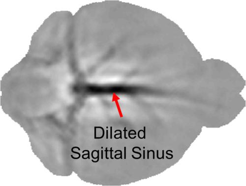
Intracranial hypotension (IH) is a pathophysiological condition of reduced intracranial pressure caused by low cerebrospinal fluid (CSF) volume due to dural injuries from lumbar puncture, surgery, or trauma. Understanding the prognosis of IH in small animal models is important to gain insights on the complications associated with it such as orthostatic headache, cerebral venous thrombosis, coma, and so forth. Photoacoustic tomography (PAT) offers a novel and cost‐effective way to perceive and detect IH in small animal models. In this study, a pulsed laser diode (PLD)‐based PAT imaging system was used to examine the changes in the venous sinuses of the rat brain due to IH, induced through CSF extraction. After the CSF extraction, an increase in the sagittal sinus area by ~30% and width by 40% ± 5% was observed. These results provide supportive evidence that the PLD‐PAT can be employed for detecting changes in sagittal sinus due to IH in rat model.
中文翻译:

使用脉冲激光二极管光声层析成像技术在体内检测小动物颅内低血压引起的静脉窦扩张。
颅内低血压(IH)是由于腰椎穿刺,手术或外伤引起的硬脑膜损伤而导致脑脊液(CSF)体积低导致颅内压降低的病理生理状况。了解小动物模型中IH的预后对于深入了解与其相关的并发症(例如直立性头痛,脑静脉血栓形成,昏迷等)很重要。光声层析成像(PAT)提供了一种新颖且经济高效的方式来感知和检测小型动物模型中的IH。在这项研究中,使用了基于脉冲激光二极管(PLD)的PAT成像系统来检查由CSF提取引起的IH引起的大鼠脑静脉窦的变化。CSF提取后,观察到矢状窦面积增加了约30%,宽度增加了40%±5%。
更新日期:2020-03-20
中文翻译:
使用脉冲激光二极管光声层析成像技术在体内检测小动物颅内低血压引起的静脉窦扩张。
颅内低血压(IH)是由于腰椎穿刺,手术或外伤引起的硬脑膜损伤而导致脑脊液(CSF)体积低导致颅内压降低的病理生理状况。了解小动物模型中IH的预后对于深入了解与其相关的并发症(例如直立性头痛,脑静脉血栓形成,昏迷等)很重要。光声层析成像(PAT)提供了一种新颖且经济高效的方式来感知和检测小型动物模型中的IH。在这项研究中,使用了基于脉冲激光二极管(PLD)的PAT成像系统来检查由CSF提取引起的IH引起的大鼠脑静脉窦的变化。CSF提取后,观察到矢状窦面积增加了约30%,宽度增加了40%±5%。


 京公网安备 11010802027423号