当前位置:
X-MOL 学术
›
WIREs Mech. Dis.
›
论文详情
Our official English website, www.x-mol.net, welcomes your feedback! (Note: you will need to create a separate account there.)
Structural neuroimaging of the altered brain stemming from pediatric and adolescent hearing loss-Scientific and clinical challenges.
WIREs Mechanisms of Disease ( IF 3.1 ) Pub Date : 2019-12-04 , DOI: 10.1002/wsbm.1469 J Tilak Ratnanather 1, 2, 3
WIREs Mechanisms of Disease ( IF 3.1 ) Pub Date : 2019-12-04 , DOI: 10.1002/wsbm.1469 J Tilak Ratnanather 1, 2, 3
Affiliation
|
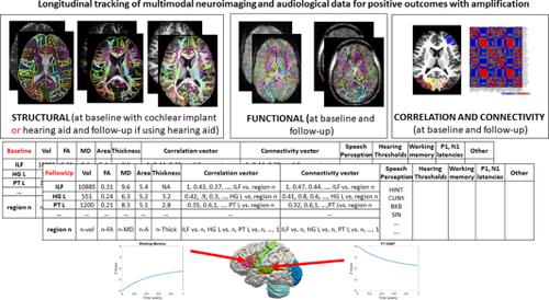
There has been a spurt in structural neuroimaging studies of the effect of hearing loss on the brain. Specifically, magnetic resonance imaging (MRI) and diffusion tensor imaging (DTI) technologies provide an opportunity to quantify changes in gray and white matter structures at the macroscopic scale. To date, there have been 32 MRI and 23 DTI studies that have analyzed structural differences accruing from pre‐ or peri‐lingual pediatric hearing loss with congenital or early onset etiology and postlingual hearing loss in pre‐to‐late adolescence. Additionally, there have been 15 prospective clinical structural neuroimaging studies of children and adolescents being evaluated for cochlear implants. The results of the 70 studies are summarized in two figures and three tables. Plastic changes in the brain are seen to be multifocal rather than diffuse, that is, differences are consistent across regions implicated in the hearing, speech and language networks regardless of modes of communication and amplification. Structures in that play an important role in cognition are affected to a lesser extent. A limitation of these studies is the emphasis on volumetric measures and on homogeneous groups of subjects with hearing loss. It is suggested that additional measures of morphometry and connectivity could contribute to a greater understanding of the effect of hearing loss on the brain. Then an interpretation of the observed macroscopic structural differences is given. This is followed by discussion of how structural imaging can be combined with functional imaging to provide biomarkers for longitudinal tracking of amplification.
中文翻译:

小儿和青少年听力损失引起的大脑改变的结构神经影像学-科学和临床挑战。
关于听力损失对大脑的影响的结构神经影像学研究激增。具体而言,磁共振成像(MRI)和扩散张量成像(DTI)技术提供了在宏观尺度上量化灰色和白色物质结构变化的机会。迄今为止,已有32项MRI和23项DTI研究分析了结构性差异,这些结构差异是由于先天或早发的儿科听力损失伴有先天性或早发性病因,以及青春期前到晚期的舌后听力损失所致。此外,已有15项针对儿童和青少年的前瞻性临床结构神经影像学研究正在接受耳蜗植入物的评估。这70项研究的结果总结在两个数字和三个表格中。大脑中的塑性变化被认为是多灶性而非扩散性的,也就是说,无论交流和放大方式如何,在涉及听觉,语音和语言网络的各个区域之间的差异都是一致的。在认知中起重要作用的结构受到的影响较小。这些研究的局限性在于对体积测量的重视以及对听力损失受试者的同类人群的重视。有人提出,形态测量学和连通性的其他措施可能有助于更好地了解听力损失对大脑的影响。然后给出了观察到的宏观结构差异的解释。接下来是关于如何将结构成像与功能成像结合以提供生物标记物用于纵向追踪扩增的讨论。语音和语言网络,无论沟通和放大模式如何。在认知中起重要作用的结构受到的影响较小。这些研究的局限性在于对体积测量的重视以及对听力损失受试者的同类人群的重视。有人提出,形态测量学和连通性的其他措施可能有助于更好地了解听力损失对大脑的影响。然后给出了观察到的宏观结构差异的解释。接下来是关于如何将结构成像与功能成像结合以提供生物标记物用于纵向追踪扩增的讨论。语音和语言网络,无论沟通和放大模式如何。在认知中起重要作用的结构受到的影响较小。这些研究的局限性在于对体积测量的重视以及对听力损失受试者的同类人群的重视。有人提出,形态测量学和连通性的其他措施可能有助于更好地了解听力损失对大脑的影响。然后给出了观察到的宏观结构差异的解释。接下来是关于如何将结构成像与功能成像结合以提供生物标记物用于纵向追踪扩增的讨论。在认知中起重要作用的结构受到的影响较小。这些研究的局限性在于对体积测量的重视以及对听力损失受试者的同类人群的重视。有人提出,形态测量学和连通性的其他措施可能有助于更好地了解听力损失对大脑的影响。然后给出了观察到的宏观结构差异的解释。接下来是关于如何将结构成像与功能成像结合以提供生物标记物用于纵向追踪扩增的讨论。在认知中起重要作用的结构受到的影响较小。这些研究的局限性在于对体积测量的重视以及对听力损失受试者的同类人群的重视。有人提出,形态测量学和连通性的其他措施可能有助于更好地了解听力损失对大脑的影响。然后给出了观察到的宏观结构差异的解释。接下来是关于如何将结构成像与功能成像结合以提供生物标记物用于纵向追踪扩增的讨论。有人提出,形态测量学和连通性的其他措施可能有助于更好地了解听力损失对大脑的影响。然后给出了观察到的宏观结构差异的解释。接下来是关于如何将结构成像与功能成像结合以提供生物标记物用于纵向追踪扩增的讨论。有人提出,形态测量学和连通性的其他措施可能有助于更好地了解听力损失对大脑的影响。然后给出了观察到的宏观结构差异的解释。接下来是关于如何将结构成像与功能成像结合以提供生物标记物用于纵向追踪扩增的讨论。
更新日期:2019-12-04
中文翻译:
小儿和青少年听力损失引起的大脑改变的结构神经影像学-科学和临床挑战。
关于听力损失对大脑的影响的结构神经影像学研究激增。具体而言,磁共振成像(MRI)和扩散张量成像(DTI)技术提供了在宏观尺度上量化灰色和白色物质结构变化的机会。迄今为止,已有32项MRI和23项DTI研究分析了结构性差异,这些结构差异是由于先天或早发的儿科听力损失伴有先天性或早发性病因,以及青春期前到晚期的舌后听力损失所致。此外,已有15项针对儿童和青少年的前瞻性临床结构神经影像学研究正在接受耳蜗植入物的评估。这70项研究的结果总结在两个数字和三个表格中。大脑中的塑性变化被认为是多灶性而非扩散性的,也就是说,无论交流和放大方式如何,在涉及听觉,语音和语言网络的各个区域之间的差异都是一致的。在认知中起重要作用的结构受到的影响较小。这些研究的局限性在于对体积测量的重视以及对听力损失受试者的同类人群的重视。有人提出,形态测量学和连通性的其他措施可能有助于更好地了解听力损失对大脑的影响。然后给出了观察到的宏观结构差异的解释。接下来是关于如何将结构成像与功能成像结合以提供生物标记物用于纵向追踪扩增的讨论。语音和语言网络,无论沟通和放大模式如何。在认知中起重要作用的结构受到的影响较小。这些研究的局限性在于对体积测量的重视以及对听力损失受试者的同类人群的重视。有人提出,形态测量学和连通性的其他措施可能有助于更好地了解听力损失对大脑的影响。然后给出了观察到的宏观结构差异的解释。接下来是关于如何将结构成像与功能成像结合以提供生物标记物用于纵向追踪扩增的讨论。语音和语言网络,无论沟通和放大模式如何。在认知中起重要作用的结构受到的影响较小。这些研究的局限性在于对体积测量的重视以及对听力损失受试者的同类人群的重视。有人提出,形态测量学和连通性的其他措施可能有助于更好地了解听力损失对大脑的影响。然后给出了观察到的宏观结构差异的解释。接下来是关于如何将结构成像与功能成像结合以提供生物标记物用于纵向追踪扩增的讨论。在认知中起重要作用的结构受到的影响较小。这些研究的局限性在于对体积测量的重视以及对听力损失受试者的同类人群的重视。有人提出,形态测量学和连通性的其他措施可能有助于更好地了解听力损失对大脑的影响。然后给出了观察到的宏观结构差异的解释。接下来是关于如何将结构成像与功能成像结合以提供生物标记物用于纵向追踪扩增的讨论。在认知中起重要作用的结构受到的影响较小。这些研究的局限性在于对体积测量的重视以及对听力损失受试者的同类人群的重视。有人提出,形态测量学和连通性的其他措施可能有助于更好地了解听力损失对大脑的影响。然后给出了观察到的宏观结构差异的解释。接下来是关于如何将结构成像与功能成像结合以提供生物标记物用于纵向追踪扩增的讨论。有人提出,形态测量学和连通性的其他措施可能有助于更好地了解听力损失对大脑的影响。然后给出了观察到的宏观结构差异的解释。接下来是关于如何将结构成像与功能成像结合以提供生物标记物用于纵向追踪扩增的讨论。有人提出,形态测量学和连通性的其他措施可能有助于更好地了解听力损失对大脑的影响。然后给出了观察到的宏观结构差异的解释。接下来是关于如何将结构成像与功能成像结合以提供生物标记物用于纵向追踪扩增的讨论。


 京公网安备 11010802027423号