当前位置:
X-MOL 学术
›
Microsc. Res. Tech.
›
论文详情
Our official English website, www.x-mol.net, welcomes your feedback! (Note: you will need to create a separate account there.)
Image mean square displacement to study the lateral mobility of Angiotensin II type 1 and Endothelin 1 type A receptors on living cells
Microscopy Research and Technique ( IF 2.5 ) Pub Date : 2019-12-13 , DOI: 10.1002/jemt.23425 Nadir Planes 1, 2 , Patrick P M L Vanderheyden 3 , Enrico Gratton 4 , Catherina Caballero-George 1
Microscopy Research and Technique ( IF 2.5 ) Pub Date : 2019-12-13 , DOI: 10.1002/jemt.23425 Nadir Planes 1, 2 , Patrick P M L Vanderheyden 3 , Enrico Gratton 4 , Catherina Caballero-George 1
Affiliation
|
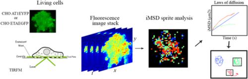
The lateral mobility of membrane receptors provides insights into the molecular interactions of protein binding and the complex dynamic plasma membrane. The image mean square displacement (iMSD) analysis is a method used to extract qualitative and quantitative information of the protein diffusion law and infers how diffusion dynamic processes may change when the cellular environment is modified. The aim of the study was to describe the membrane diffusing properties of two G‐protein‐coupled receptors namely Angiotensin II type 1 (AT1) and Endothelin 1 type A (ETA) receptors and their corresponding receptor–ligand complexes in living cells using total internal reflection fluorescent microscopy and iMSD analysis. This study showed that both AT1 and ETA receptors displayed a mix of three modes of diffusion: free, confined, and partially confined. The confined mode was the predominant at the plasma membrane of living cells and was not affected by ligand binding. However, the local diffusivity and the confinement zone of AT1 receptors were reduced by the binding of its antagonist losartan, and the long‐range diffusion with the local diffusivity coefficient of ETA receptors was reduced upon exposure to its antagonist BQ123. To the best of our knowledge, this is the first study addressing the protein diffusion laws of these two receptors on living cells using total internal reflection fluorescence microscopy and iMSD.
中文翻译:

图像均方位移以研究血管紧张素II 1型和内皮素1 A型受体在活细胞上的横向迁移
膜受体的横向迁移性提供了对蛋白质结合和复杂动态质膜的分子相互作用的见解。图像均方位移(iMSD)分析是一种用于提取蛋白质扩散定律的定性和定量信息,并推断在修改细胞环境时扩散动态过程可能如何变化的方法。这项研究的目的是描述两种G蛋白偶联受体,即血管紧张素II 1型(AT 1)和内皮素1 A型(ET A)受体及其在活细胞中相应的受体-配体复合物的膜扩散特性。全内反射荧光显微镜和iMSD分析。这项研究表明AT 1和ET甲受体显示的扩散的三种模式的混合:免费,密闭,和部分密闭。受限模式主要存在于活细胞的质膜上,不受配体结合的影响。然而,AT 1受体的拮抗剂洛沙坦的结合降低了AT 1受体的局部扩散和限制区,暴露于其拮抗剂BQ123时,ET A受体的局部扩散系数的远距离扩散降低了。据我们所知,这是首次使用全内反射荧光显微镜和iMSD研究这两个受体在活细胞上的蛋白质扩散规律的研究。
更新日期:2019-12-13
中文翻译:
图像均方位移以研究血管紧张素II 1型和内皮素1 A型受体在活细胞上的横向迁移
膜受体的横向迁移性提供了对蛋白质结合和复杂动态质膜的分子相互作用的见解。图像均方位移(iMSD)分析是一种用于提取蛋白质扩散定律的定性和定量信息,并推断在修改细胞环境时扩散动态过程可能如何变化的方法。这项研究的目的是描述两种G蛋白偶联受体,即血管紧张素II 1型(AT 1)和内皮素1 A型(ET A)受体及其在活细胞中相应的受体-配体复合物的膜扩散特性。全内反射荧光显微镜和iMSD分析。这项研究表明AT 1和ET甲受体显示的扩散的三种模式的混合:免费,密闭,和部分密闭。受限模式主要存在于活细胞的质膜上,不受配体结合的影响。然而,AT 1受体的拮抗剂洛沙坦的结合降低了AT 1受体的局部扩散和限制区,暴露于其拮抗剂BQ123时,ET A受体的局部扩散系数的远距离扩散降低了。据我们所知,这是首次使用全内反射荧光显微镜和iMSD研究这两个受体在活细胞上的蛋白质扩散规律的研究。


 京公网安备 11010802027423号