当前位置:
X-MOL 学术
›
Dent. Mater.
›
论文详情
Our official English website, www.x-mol.net, welcomes your feedback! (Note: you will need to create a separate account there.)
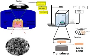
Minimally invasive high-intensity focused ultrasound (HIFU) improves dentine remineralization with hydroxyapatite nanorods.
Dental Materials ( IF 5 ) Pub Date : 2020-01-31 , DOI: 10.1016/j.dental.2020.01.005 Umer Daood 1 , A S Fawzy 2
Dental Materials ( IF 5 ) Pub Date : 2020-01-31 , DOI: 10.1016/j.dental.2020.01.005 Umer Daood 1 , A S Fawzy 2
Affiliation
|
OBJECTIVE
The aim is to investigate the potential significance of combining minimally invasive high-intensity focused ultrasound (HIFU) with hydroxyapatite (HA) nanorods treatment for the remineralization of demineralized coronal dentine-matrix.
METHODS
HA having nanorods structure were synthetized using ultrasonication with precipitation method. HA nanorods were characterized by TEM for average-size/shape. Following phosphoric acid demineralization, dentine specimens were treated with HA-nanorods with/without subsequent HIFU exposure for 5 s, 10 s and 20 s then stored in artificial saliva for 1-month. Dentine specimens were characterized using different SEM and Raman spectroscopic techniques. In addition, the biochemical stability and HA-nanorods were examined using ATR-FTIR to observe attachment of nanoparticles. Also, surface nanoindentation properties were evaluated using AFM in tapping-mode.
RESULTS
HA-nanorods displayed well-defined, homogenous plate-like nanostructure. TEM revealed intact collagen-fibrils network structure with high density due to obliteration of interfibrillar spaces with clear evidence of remineralization in combined HA/HIFU treatment. With HA-nanorods treatment collagen-network structure was visible, consisting of fibrils interlaced into a compact pattern with evidence of minerals deposition. AFM investigation revealed clear mineral formation with the increase of HIFU exposure time. Bands associated with inorganic phase dominate well in HIFU exposed specimens with PO stretching within dentine mineral identified at 960 cm-1. Characteristic dentine structure for control and HIFU 20 s specimens is reflected as oscillatory mean Amide-I intensity with measurement giving a precise sinusoidal response of polarization angle β within dentinal tissue. Nanoindentation testing showed a gradual significant increase in elastic-modulus with the increase in HIFU exposure time after 1-month storage. FTIR spectrum of the HIFU exposed dentine displayed bands at 1650 cm-1, 1580 cm-1 and 1510 cm-1 that can be attributed to Amide-I, II and III.
SIGNIFICANCE
The synergetic effect of HIFU exposure on remineralization potential of demineralized dentine-matrix following nano-hydroxyapatite treatment was revealed. This synergetic effect is dependent on HIFU exposure time.
中文翻译:

微创高强度聚焦超声(HIFU)使用羟基磷灰石纳米棒改善牙本质的再矿化作用。
目的目的是研究将微创高强度聚焦超声(HIFU)与羟基磷灰石(HA)纳米棒相结合对脱矿质冠状牙本质基质再矿化的潜在意义。方法采用沉淀法超声处理合成纳米棒结构的HA。HA纳米棒通过TEM表征平均尺寸/形状。磷酸脱矿质后,将牙本质标本用HA-nanorods处理,有/无随后HIFU暴露5 s,10 s和20 s,然后在人造唾液中保存1个月。使用不同的SEM和拉曼光谱技术对牙本质标本进行表征。另外,使用ATR-FTIR检查了生化稳定性和HA-纳米脚,以观察纳米颗粒的附着。也,使用AFM在攻丝模式下评估表面纳米压痕性能。结果HA-nanorods显示出清晰,均质的板状纳米结构。透射电镜显示完整的胶原-纤维网络结构由于纤维间间隙的消除而具有高密度,并有明确的证据表明在HA / HIFU联合治疗中会再矿化。用HA-nanorods处理后,可见胶原蛋白网络结构,其由交织成紧密模式的原纤维组成,具有矿物质沉积的迹象。原子力显微镜调查显示,随着HIFU暴露时间的增加,清楚的矿物形成。在HIFU暴露的标本中,与无机相相关的谱带占主导地位,在960 cm-1处发现的牙本质矿物中有PO拉伸。对照和HIFU 20 s标本的特征性牙本质结构反映为振荡平均Amide-I强度,并通过测量给出了牙本质组织内极化角β的精确正弦响应。纳米压痕测试显示,在储存1个月后,弹性模量随着HIFU暴露时间的增加而逐渐增加。HIFU暴露的牙本质的FTIR光谱显示了1650 cm-1、1580 cm-1和1510 cm-1的条带,这可以归因于酰胺I,II和III。意义揭示了HIFU暴露对纳米羟基磷灰石处理后脱矿牙本质基质再矿化潜力的协同作用。这种协同作用取决于HIFU的暴露时间。纳米压痕测试显示,在储存1个月后,弹性模量随着HIFU暴露时间的增加而逐渐增加。HIFU暴露的牙本质的FTIR光谱显示了1650 cm-1、1580 cm-1和1510 cm-1的条带,这可以归因于酰胺I,II和III。意义揭示了HIFU暴露对纳米羟基磷灰石处理后脱矿牙本质基质再矿化潜力的协同作用。这种协同作用取决于HIFU的暴露时间。纳米压痕测试显示,在储存1个月后,弹性模量随着HIFU暴露时间的增加而逐渐增加。HIFU暴露的牙本质的FTIR光谱显示了1650 cm-1、1580 cm-1和1510 cm-1的条带,这可以归因于酰胺I,II和III。意义揭示了HIFU暴露对纳米羟基磷灰石处理后脱矿牙本质基质再矿化潜力的协同作用。这种协同作用取决于HIFU的暴露时间。意义揭示了HIFU暴露对纳米羟基磷灰石处理后脱矿牙本质基质再矿化潜力的协同作用。这种协同作用取决于HIFU的暴露时间。意义揭示了HIFU暴露对纳米羟基磷灰石处理后脱矿牙本质基质再矿化潜力的协同作用。这种协同作用取决于HIFU的暴露时间。
更新日期:2020-01-31
中文翻译:
微创高强度聚焦超声(HIFU)使用羟基磷灰石纳米棒改善牙本质的再矿化作用。
目的目的是研究将微创高强度聚焦超声(HIFU)与羟基磷灰石(HA)纳米棒相结合对脱矿质冠状牙本质基质再矿化的潜在意义。方法采用沉淀法超声处理合成纳米棒结构的HA。HA纳米棒通过TEM表征平均尺寸/形状。磷酸脱矿质后,将牙本质标本用HA-nanorods处理,有/无随后HIFU暴露5 s,10 s和20 s,然后在人造唾液中保存1个月。使用不同的SEM和拉曼光谱技术对牙本质标本进行表征。另外,使用ATR-FTIR检查了生化稳定性和HA-纳米脚,以观察纳米颗粒的附着。也,使用AFM在攻丝模式下评估表面纳米压痕性能。结果HA-nanorods显示出清晰,均质的板状纳米结构。透射电镜显示完整的胶原-纤维网络结构由于纤维间间隙的消除而具有高密度,并有明确的证据表明在HA / HIFU联合治疗中会再矿化。用HA-nanorods处理后,可见胶原蛋白网络结构,其由交织成紧密模式的原纤维组成,具有矿物质沉积的迹象。原子力显微镜调查显示,随着HIFU暴露时间的增加,清楚的矿物形成。在HIFU暴露的标本中,与无机相相关的谱带占主导地位,在960 cm-1处发现的牙本质矿物中有PO拉伸。对照和HIFU 20 s标本的特征性牙本质结构反映为振荡平均Amide-I强度,并通过测量给出了牙本质组织内极化角β的精确正弦响应。纳米压痕测试显示,在储存1个月后,弹性模量随着HIFU暴露时间的增加而逐渐增加。HIFU暴露的牙本质的FTIR光谱显示了1650 cm-1、1580 cm-1和1510 cm-1的条带,这可以归因于酰胺I,II和III。意义揭示了HIFU暴露对纳米羟基磷灰石处理后脱矿牙本质基质再矿化潜力的协同作用。这种协同作用取决于HIFU的暴露时间。纳米压痕测试显示,在储存1个月后,弹性模量随着HIFU暴露时间的增加而逐渐增加。HIFU暴露的牙本质的FTIR光谱显示了1650 cm-1、1580 cm-1和1510 cm-1的条带,这可以归因于酰胺I,II和III。意义揭示了HIFU暴露对纳米羟基磷灰石处理后脱矿牙本质基质再矿化潜力的协同作用。这种协同作用取决于HIFU的暴露时间。纳米压痕测试显示,在储存1个月后,弹性模量随着HIFU暴露时间的增加而逐渐增加。HIFU暴露的牙本质的FTIR光谱显示了1650 cm-1、1580 cm-1和1510 cm-1的条带,这可以归因于酰胺I,II和III。意义揭示了HIFU暴露对纳米羟基磷灰石处理后脱矿牙本质基质再矿化潜力的协同作用。这种协同作用取决于HIFU的暴露时间。意义揭示了HIFU暴露对纳米羟基磷灰石处理后脱矿牙本质基质再矿化潜力的协同作用。这种协同作用取决于HIFU的暴露时间。意义揭示了HIFU暴露对纳米羟基磷灰石处理后脱矿牙本质基质再矿化潜力的协同作用。这种协同作用取决于HIFU的暴露时间。


 京公网安备 11010802027423号