当前位置:
X-MOL 学术
›
Electroanalysis
›
论文详情
Our official English website, www.x-mol.net, welcomes your feedback! (Note: you will need to create a separate account there.)
Electroanalytical Method for Quantification of Hepatocellular Carcinoma Cells as Charge Transport Barriers in Culture Media
Electroanalysis ( IF 3 ) Pub Date : 2020-01-28 , DOI: 10.1002/elan.201900553 Juhi Jaiswal 1 , Marshal Dhayal 1
Electroanalysis ( IF 3 ) Pub Date : 2020-01-28 , DOI: 10.1002/elan.201900553 Juhi Jaiswal 1 , Marshal Dhayal 1
Affiliation
|
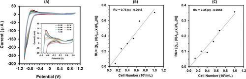
Here we report a strategy in which electroanalytical method was used to quantify human hepatic carcinoma cell (HepG2) density in culture media and their interaction with biomolecules. The cyclic voltammogram response of vitamin and amino acids redox mediators containing culture media had shown distinct oxidation peak at −0.63 V along with a low intensity peak at +0.67 V. Both oxidation and reduction peak current of culture media were gradually decreased with an increase in cell number indicating their role as charge transport barrier at electrode surface. The difference between cathodic and anodic peak potential was also decreased with the addition of cells. The oxidation peak disappeared in CV response, with the addition of optimum cell number in culture media, indicating the adsorption of redox mediators at cell surface. CV response of fetal bovine serum (FBS) containing cell suspension showed presence of reduction and oxidation peaks of culture media in CV. This indicates stronger possibility of binding of serum proteins with cells and release of redox mediators in culture media. The chemical interactions of cells with FBS was further confirmed by the FTIR and UV‐Vis spectroscopy. The viability, adhesion and proliferation responses of the cells were found to be normal. The reported electroanalytical method may be applied in the future for rapid quantification of cell density and confirmation of interactions among cells and biomolecules.
中文翻译:

定量分析肝细胞癌细胞作为培养基中电荷传输屏障的电分析方法
在这里,我们报告一种策略,其中电分析方法用于量化培养基中人类肝癌细胞(HepG2)的密度及其与生物分子的相互作用。含有培养基的维生素和氨基酸氧化还原介体的循环伏安图响应显示出在-0.63 V处有明显的氧化峰,在+0.67 V处有较低的强度峰。随着Cd的增加,培养基的氧化和还原峰电流逐渐降低。表示它们作为电极表面电荷传输屏障的作用的电池数目。阴极和阳极峰值电位之间的差异也随着添加细胞而减小。在培养基中添加最佳细胞数后,CV反应中的氧化峰消失,表明氧化还原介质在细胞表面的吸附。含有细胞悬浮液的胎牛血清(FBS)的CV响应显示在CV中存在培养基的还原和氧化峰。这表明血清蛋白与细胞结合以及氧化还原介体在培养基中释放的可能性更高。FTIR和UV-Vis光谱进一步证实了细胞与FBS的化学相互作用。发现细胞的活力,粘附和增殖反应是正常的。所报道的电分析方法可在将来用于细胞密度的快速定量和细胞与生物分子之间相互作用的确认。这表明血清蛋白与细胞结合以及氧化还原介体在培养基中释放的可能性更高。FTIR和UV-Vis光谱进一步证实了细胞与FBS的化学相互作用。发现细胞的活力,粘附和增殖反应是正常的。所报道的电分析方法可在将来用于细胞密度的快速定量和细胞与生物分子之间相互作用的确认。这表明血清蛋白与细胞结合以及氧化还原介体在培养基中释放的可能性更高。FTIR和UV-Vis光谱进一步证实了细胞与FBS的化学相互作用。发现细胞的活力,粘附和增殖反应是正常的。所报道的电分析方法可在将来用于细胞密度的快速定量和细胞与生物分子之间相互作用的确认。
更新日期:2020-01-28
中文翻译:
定量分析肝细胞癌细胞作为培养基中电荷传输屏障的电分析方法
在这里,我们报告一种策略,其中电分析方法用于量化培养基中人类肝癌细胞(HepG2)的密度及其与生物分子的相互作用。含有培养基的维生素和氨基酸氧化还原介体的循环伏安图响应显示出在-0.63 V处有明显的氧化峰,在+0.67 V处有较低的强度峰。随着Cd的增加,培养基的氧化和还原峰电流逐渐降低。表示它们作为电极表面电荷传输屏障的作用的电池数目。阴极和阳极峰值电位之间的差异也随着添加细胞而减小。在培养基中添加最佳细胞数后,CV反应中的氧化峰消失,表明氧化还原介质在细胞表面的吸附。含有细胞悬浮液的胎牛血清(FBS)的CV响应显示在CV中存在培养基的还原和氧化峰。这表明血清蛋白与细胞结合以及氧化还原介体在培养基中释放的可能性更高。FTIR和UV-Vis光谱进一步证实了细胞与FBS的化学相互作用。发现细胞的活力,粘附和增殖反应是正常的。所报道的电分析方法可在将来用于细胞密度的快速定量和细胞与生物分子之间相互作用的确认。这表明血清蛋白与细胞结合以及氧化还原介体在培养基中释放的可能性更高。FTIR和UV-Vis光谱进一步证实了细胞与FBS的化学相互作用。发现细胞的活力,粘附和增殖反应是正常的。所报道的电分析方法可在将来用于细胞密度的快速定量和细胞与生物分子之间相互作用的确认。这表明血清蛋白与细胞结合以及氧化还原介体在培养基中释放的可能性更高。FTIR和UV-Vis光谱进一步证实了细胞与FBS的化学相互作用。发现细胞的活力,粘附和增殖反应是正常的。所报道的电分析方法可在将来用于细胞密度的快速定量和细胞与生物分子之间相互作用的确认。


 京公网安备 11010802027423号