当前位置:
X-MOL 学术
›
Modern Pathol.
›
论文详情
Our official English website, www.x-mol.net, welcomes your feedback! (Note: you will need to create a separate account there.)
Intense basolateral membrane staining indicates HER2 positivity in invasive micropapillary breast carcinoma.
Modern Pathology ( IF 7.5 ) Pub Date : 2020-01-23 , DOI: 10.1038/s41379-020-0461-z Shuling Zhou 1, 2 , Fei Yang 1, 2 , Qianming Bai 1, 2 , Anqi Li 1, 2 , Ming Li 1, 2 , Siyuan Zhong 1, 2 , Hong Lv 1, 2 , Ruohong Shui 1, 2 , Xiaoyu Tu 1, 2 , Rui Bi 1, 2 , Xiaoli Xu 1, 2 , Yufan Cheng 1, 2 , Baohua Yu 1, 2 , Shaoxian Tang 1, 2 , Xiangjie Sun 1, 2 , Xiaoyan Zhou 1, 2 , Wentao Yang 1, 2
Modern Pathology ( IF 7.5 ) Pub Date : 2020-01-23 , DOI: 10.1038/s41379-020-0461-z Shuling Zhou 1, 2 , Fei Yang 1, 2 , Qianming Bai 1, 2 , Anqi Li 1, 2 , Ming Li 1, 2 , Siyuan Zhong 1, 2 , Hong Lv 1, 2 , Ruohong Shui 1, 2 , Xiaoyu Tu 1, 2 , Rui Bi 1, 2 , Xiaoli Xu 1, 2 , Yufan Cheng 1, 2 , Baohua Yu 1, 2 , Shaoxian Tang 1, 2 , Xiangjie Sun 1, 2 , Xiaoyan Zhou 1, 2 , Wentao Yang 1, 2
Affiliation
|
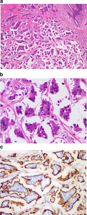
Invasive micropapillary carcinoma is characterized by the inside-out growth of tumor clusters and displays incomplete membrane immunostaining of HER2. According to the 2018 American Society of Clinical Oncology and the College of American Pathologists (ASCO/CAP) HER2-testing recommendation, moderate to intense but incomplete staining could be scored as immunohistochemical 2+. Furthermore, the criteria of immunohistochemical 3+ for this staining pattern are not mentioned. One hundred and forty-seven cases of invasive micropapillary carcinoma with moderate-to-intense HER2 immunostaining were enrolled. Invasive micropapillary carcinoma components of all cases were scored as immunohistochemical 2+ based on the 2018 ASCO/CAP recommendation. The invasive micropapillary carcinoma component varied from 10% to 100% (mean, 80%). Invasive micropapillary carcinoma components of all 147 tumors exhibited reversed polarity and incomplete basolateral HER2 membrane staining. One hundred and seventeen of the tumors (80%, 117/147) had moderate staining, and 38 (32%, 38/117) showed HER2 gene amplification by fluorescence in-situ hybridization. HER2 gene was amplified in all the remaining 30 tumors (20%, 30/147) that exhibited intense basolateral membrane staining. Besides, average HER2 signals per cell and ratio of HER2/CEP17 were significantly higher in the intense-staining tumors compared with the moderate-staining tumors (p < 0.0001). Follow-up data were available for 140 patients. None of the patients were died. The follow-up time ranged from 1 month to 99 months (median, 57 months). Thirteen (9%, 13/140) patients exhibited disease progression (recurrence or metastasis). HER2 gene amplification was correlated inversely with estrogen receptor (p = 0.000) and progesterone receptor (p = 0.000) expression, and positively with histological grade (p = 0.003) and disease progression (p = 0.000). Invasive micropapillary carcinoma with intense clear linear basolateral membrane immunostaining indicates HER2 positivity, even if the staining is incomplete. They should be classified as immunohistochemical 3+ rather than immunohistochemical 2+, which would avoid further fluorescence in-situ hybridization-testing procedure and greatly save the related time, labor, and financial costs. Ultimately, ensure all patients with HER2 gene amplification obtain effective targeted therapy in time.
中文翻译:

强烈的基底外侧膜染色表明浸润性微乳头状乳腺癌中的 HER2 阳性。
浸润性微乳头状癌的特征是肿瘤簇由内向外生长,并且显示 HER2 的膜免疫染色不完整。根据 2018 年美国临床肿瘤学会和美国病理学家学会 (ASCO/CAP) HER2 检测建议,中度至强烈但不完全染色可评分为免疫组化 2+。此外,未提及此染色模式的免疫组化 3+ 标准。纳入了 147 例具有中度至强烈 HER2 免疫染色的浸润性微乳头状癌。所有病例的浸润性微乳头状癌成分均根据2018年ASCO/CAP推荐评分为免疫组化2+。浸润性微乳头状癌成分从 10% 到 100% 不等(平均 80%)。所有 147 个肿瘤的浸润性微乳头状癌成分均表现出极性反转和基底外侧 HER2 膜染色不完整。117 个肿瘤 (80%, 117/147) 具有中度染色,38 个 (32%, 38/117) 通过荧光原位杂交显示 HER2 基因扩增。HER2 基因在所有剩余的 30 个肿瘤(20%,30/147)中被扩增,这些肿瘤表现出强烈的基底外侧膜染色。此外,与中等染色肿瘤相比,强染色肿瘤中每个细胞的平均 HER2 信号和 HER2/CEP17 比率显着更高 (p < 0.0001)。可获得 140 名患者的随访数据。没有患者死亡。随访时间从 1 个月到 99 个月不等(中位数,57 个月)。十三名 (9%, 13/140) 患者表现出疾病进展(复发或转移)。HER2 基因扩增与雌激素受体 (p = 0.000) 和孕激素受体 (p = 0.000) 表达呈负相关,与组织学分级 (p = 0.003) 和疾病进展 (p = 0.000) 呈正相关。具有强烈清晰线性基底外侧膜免疫染色的浸润性微乳头状癌表明 HER2 阳性,即使染色不完整。它们应该被归类为免疫组化3+而不是免疫组化2+,这将避免进一步的荧光原位杂交检测程序,并大大节省相关的时间、人力和财务成本。最终确保所有HER2基因扩增的患者及时获得有效的靶向治疗。并且与组织学分级 (p = 0.003) 和疾病进展 (p = 0.000) 呈正相关。具有强烈清晰线性基底外侧膜免疫染色的浸润性微乳头状癌表明 HER2 阳性,即使染色不完整。它们应归类为免疫组化 3+ 而不是免疫组化 2+,这将避免进一步的荧光原位杂交检测程序,并大大节省相关的时间、人力和财务成本。最终确保所有HER2基因扩增的患者及时获得有效的靶向治疗。并且与组织学分级 (p = 0.003) 和疾病进展 (p = 0.000) 呈正相关。具有强烈清晰线性基底外侧膜免疫染色的浸润性微乳头状癌表明 HER2 阳性,即使染色不完整。它们应归类为免疫组化 3+ 而不是免疫组化 2+,这将避免进一步的荧光原位杂交检测程序,并大大节省相关的时间、人力和财务成本。最终确保所有HER2基因扩增的患者及时获得有效的靶向治疗。它们应归类为免疫组化 3+ 而不是免疫组化 2+,这将避免进一步的荧光原位杂交检测程序,并大大节省相关的时间、人力和财务成本。最终确保所有HER2基因扩增的患者及时获得有效的靶向治疗。它们应该被归类为免疫组化3+而不是免疫组化2+,这将避免进一步的荧光原位杂交检测程序,并大大节省相关的时间、人力和财务成本。最终确保所有HER2基因扩增的患者及时获得有效的靶向治疗。
更新日期:2020-01-24
中文翻译:
强烈的基底外侧膜染色表明浸润性微乳头状乳腺癌中的 HER2 阳性。
浸润性微乳头状癌的特征是肿瘤簇由内向外生长,并且显示 HER2 的膜免疫染色不完整。根据 2018 年美国临床肿瘤学会和美国病理学家学会 (ASCO/CAP) HER2 检测建议,中度至强烈但不完全染色可评分为免疫组化 2+。此外,未提及此染色模式的免疫组化 3+ 标准。纳入了 147 例具有中度至强烈 HER2 免疫染色的浸润性微乳头状癌。所有病例的浸润性微乳头状癌成分均根据2018年ASCO/CAP推荐评分为免疫组化2+。浸润性微乳头状癌成分从 10% 到 100% 不等(平均 80%)。所有 147 个肿瘤的浸润性微乳头状癌成分均表现出极性反转和基底外侧 HER2 膜染色不完整。117 个肿瘤 (80%, 117/147) 具有中度染色,38 个 (32%, 38/117) 通过荧光原位杂交显示 HER2 基因扩增。HER2 基因在所有剩余的 30 个肿瘤(20%,30/147)中被扩增,这些肿瘤表现出强烈的基底外侧膜染色。此外,与中等染色肿瘤相比,强染色肿瘤中每个细胞的平均 HER2 信号和 HER2/CEP17 比率显着更高 (p < 0.0001)。可获得 140 名患者的随访数据。没有患者死亡。随访时间从 1 个月到 99 个月不等(中位数,57 个月)。十三名 (9%, 13/140) 患者表现出疾病进展(复发或转移)。HER2 基因扩增与雌激素受体 (p = 0.000) 和孕激素受体 (p = 0.000) 表达呈负相关,与组织学分级 (p = 0.003) 和疾病进展 (p = 0.000) 呈正相关。具有强烈清晰线性基底外侧膜免疫染色的浸润性微乳头状癌表明 HER2 阳性,即使染色不完整。它们应该被归类为免疫组化3+而不是免疫组化2+,这将避免进一步的荧光原位杂交检测程序,并大大节省相关的时间、人力和财务成本。最终确保所有HER2基因扩增的患者及时获得有效的靶向治疗。并且与组织学分级 (p = 0.003) 和疾病进展 (p = 0.000) 呈正相关。具有强烈清晰线性基底外侧膜免疫染色的浸润性微乳头状癌表明 HER2 阳性,即使染色不完整。它们应归类为免疫组化 3+ 而不是免疫组化 2+,这将避免进一步的荧光原位杂交检测程序,并大大节省相关的时间、人力和财务成本。最终确保所有HER2基因扩增的患者及时获得有效的靶向治疗。并且与组织学分级 (p = 0.003) 和疾病进展 (p = 0.000) 呈正相关。具有强烈清晰线性基底外侧膜免疫染色的浸润性微乳头状癌表明 HER2 阳性,即使染色不完整。它们应归类为免疫组化 3+ 而不是免疫组化 2+,这将避免进一步的荧光原位杂交检测程序,并大大节省相关的时间、人力和财务成本。最终确保所有HER2基因扩增的患者及时获得有效的靶向治疗。它们应归类为免疫组化 3+ 而不是免疫组化 2+,这将避免进一步的荧光原位杂交检测程序,并大大节省相关的时间、人力和财务成本。最终确保所有HER2基因扩增的患者及时获得有效的靶向治疗。它们应该被归类为免疫组化3+而不是免疫组化2+,这将避免进一步的荧光原位杂交检测程序,并大大节省相关的时间、人力和财务成本。最终确保所有HER2基因扩增的患者及时获得有效的靶向治疗。


 京公网安备 11010802027423号