当前位置:
X-MOL 学术
›
Ultramicroscopy
›
论文详情
Our official English website, www.x-mol.net, welcomes your feedback! (Note: you will need to create a separate account there.)
Advancing characterisation with statistics from correlative electron diffraction and X-ray spectroscopy, in the scanning electron microscope
Ultramicroscopy ( IF 2.2 ) Pub Date : 2020-04-01 , DOI: 10.1016/j.ultramic.2020.112944 T P McAuliffe 1 , A Foden 1 , C Bilsland 1 , D Daskalaki Mountanou 1 , D Dye 1 , T B Britton 1
Ultramicroscopy ( IF 2.2 ) Pub Date : 2020-04-01 , DOI: 10.1016/j.ultramic.2020.112944 T P McAuliffe 1 , A Foden 1 , C Bilsland 1 , D Daskalaki Mountanou 1 , D Dye 1 , T B Britton 1
Affiliation
|
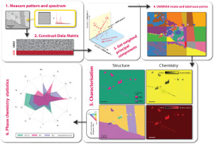
The routine and unique determination of minor phases in microstructures is critical to materials science. In metallurgy alone, applications include alloy and process development and the understanding of degradation in service. We develop a correlative method, exploring superalloy microstructures, which are examined in the scanning electron microscope (SEM) using simultaneous energy dispersive X-ray spectroscopy (EDS) and electron backscatter diffraction (EBSD). This is performed at an appropriate length scale for characterisation of carbide phases' shape, size, location, and distribution. EDS and EBSD data are generated using two different physical processes, but each provide a signature of the material interacting with the incoming electron beam. Recent advances in post-processing, driven by 'big data' approaches, include use of principal component analysis (PCA). Components are subsequently characterised to assign labels to a mapped region. To provide physically meaningful signals, the principal components may be rotated to control the distribution of variance. In this work, we develop this method further through a weighted PCA approach. We use the EDS and EBSD signals concurrently, thereby labelling each region using both EDS (chemistry) and EBSD (crystal structure) information. This provides a new method of amplifying signal-to-noise for very small phases in mapped regions, especially where the EDS or EBSD signal is not unique enough alone for classification.
中文翻译:

在扫描电子显微镜中使用相关电子衍射和 X 射线光谱的统计数据进行表征
微观结构中次要相的常规和独特测定对于材料科学至关重要。仅在冶金领域,应用就包括合金和工艺开发以及对服务退化的理解。我们开发了一种相关方法,探索高温合金微观结构,使用同步能量色散 X 射线光谱 (EDS) 和电子背散射衍射 (EBSD) 在扫描电子显微镜 (SEM) 中进行检查。这是在适当的长度尺度上进行的,用于表征碳化物相的形状、尺寸、位置和分布。EDS 和 EBSD 数据是使用两种不同的物理过程生成的,但每个过程都提供了材料与入射电子束相互作用的特征。由“大数据”方法驱动的后处理的最新进展,包括使用主成分分析 (PCA)。随后对组件进行表征以将标签分配给映射区域。为了提供物理上有意义的信号,可以旋转主成分以控制方差的分布。在这项工作中,我们通过加权 PCA 方法进一步开发了这种方法。我们同时使用 EDS 和 EBSD 信号,从而使用 EDS(化学)和 EBSD(晶体结构)信息标记每个区域。这为映射区域中非常小的相位提供了一种放大信噪比的新方法,尤其是在 EDS 或 EBSD 信号单独不足以进行分类的情况下。可以旋转主成分以控制方差的分布。在这项工作中,我们通过加权 PCA 方法进一步开发了这种方法。我们同时使用 EDS 和 EBSD 信号,从而使用 EDS(化学)和 EBSD(晶体结构)信息标记每个区域。这为映射区域中非常小的相位提供了一种放大信噪比的新方法,尤其是在 EDS 或 EBSD 信号单独不足以进行分类的情况下。可以旋转主成分以控制方差的分布。在这项工作中,我们通过加权 PCA 方法进一步开发了这种方法。我们同时使用 EDS 和 EBSD 信号,从而使用 EDS(化学)和 EBSD(晶体结构)信息标记每个区域。这为映射区域中非常小的相位提供了一种放大信噪比的新方法,尤其是在 EDS 或 EBSD 信号单独不足以进行分类的情况下。
更新日期:2020-04-01
中文翻译:
在扫描电子显微镜中使用相关电子衍射和 X 射线光谱的统计数据进行表征
微观结构中次要相的常规和独特测定对于材料科学至关重要。仅在冶金领域,应用就包括合金和工艺开发以及对服务退化的理解。我们开发了一种相关方法,探索高温合金微观结构,使用同步能量色散 X 射线光谱 (EDS) 和电子背散射衍射 (EBSD) 在扫描电子显微镜 (SEM) 中进行检查。这是在适当的长度尺度上进行的,用于表征碳化物相的形状、尺寸、位置和分布。EDS 和 EBSD 数据是使用两种不同的物理过程生成的,但每个过程都提供了材料与入射电子束相互作用的特征。由“大数据”方法驱动的后处理的最新进展,包括使用主成分分析 (PCA)。随后对组件进行表征以将标签分配给映射区域。为了提供物理上有意义的信号,可以旋转主成分以控制方差的分布。在这项工作中,我们通过加权 PCA 方法进一步开发了这种方法。我们同时使用 EDS 和 EBSD 信号,从而使用 EDS(化学)和 EBSD(晶体结构)信息标记每个区域。这为映射区域中非常小的相位提供了一种放大信噪比的新方法,尤其是在 EDS 或 EBSD 信号单独不足以进行分类的情况下。可以旋转主成分以控制方差的分布。在这项工作中,我们通过加权 PCA 方法进一步开发了这种方法。我们同时使用 EDS 和 EBSD 信号,从而使用 EDS(化学)和 EBSD(晶体结构)信息标记每个区域。这为映射区域中非常小的相位提供了一种放大信噪比的新方法,尤其是在 EDS 或 EBSD 信号单独不足以进行分类的情况下。可以旋转主成分以控制方差的分布。在这项工作中,我们通过加权 PCA 方法进一步开发了这种方法。我们同时使用 EDS 和 EBSD 信号,从而使用 EDS(化学)和 EBSD(晶体结构)信息标记每个区域。这为映射区域中非常小的相位提供了一种放大信噪比的新方法,尤其是在 EDS 或 EBSD 信号单独不足以进行分类的情况下。


 京公网安备 11010802027423号