Theranostics ( IF 12.4 ) Pub Date : 2020-01-01 , DOI: 10.7150/thno.37623 Yuichi Abe 1, 2 , Hidekazu Hirano 3 , Hirokazu Shoji 3 , Asa Tada 1, 2 , Junko Isoyama 1, 2 , Akemi Kakudo 1, 2 , Daigo Gunji 1, 2 , Kazufumi Honda 4 , Narikazu Boku 3 , Jun Adachi 1, 2 , Takeshi Tomonaga 1, 2
|
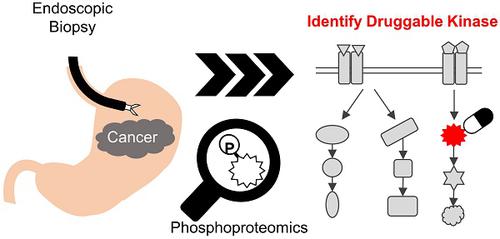
Rationale: Cancer phosphoproteomics can provide insights regarding kinases that can be targeted for therapeutic applications. Monitoring the phosphoproteomics in cancer is expected to play a key role in optimizing treatments with kinase inhibitors. Clinical phosphoproteomics in surgical tissues and patient-derived models has been studied intensively. However, the reported data may not accurately reflect the phosphosignaling status in patients due to the effect of ischemia occurring during surgery or changes in the characteristics of cancer cells when establishing the models. In contrast, endoscopic biopsies have an advantage for clinical phosphoproteomics because they can be rapidly cryo-preserved. We aimed to develop a highly sensitive method for phosphoproteomics in endoscopic biopsies of gastric cancer.
Methods: Three tumor biopsies and three normal gastric biopsies were obtained by endoscopy at one time, and subjected to our optimized phosphoproteomics. Phosphopeptides were enriched with an immobilized metal affinity chromatography, and labeled with Tandem Mass Tag reagent. Quantified phosphosites were compared between the pairs of tumor/normal biopsies within same patient. Cancer-specific activated pathways and kinases were identified by pathway enrichment analysis and kinase-substrate enrichment analysis.
Results: Our protocol enabled the identification of more than 10,000 class 1 phosphosites from endoscopic biopsies. A comparison between samples from cancer tissue and normal mucosa demonstrated differences in the phosphosignaling, including biomarkers of response to DNA damage. Finally, cancer-specific activation of DNA damage response signaling was validated by additional phosphoproteomics of other patients and western blotting of gastric cancer/normal cells.
Conclusion: In summary, our pioneering approach will facilitate more accurate clinical phosphoproteomics in endoscopic biopsies, which can be applied to monitor the activities of therapeutic kinases and, ultimately, can be a useful tool to precision medicine.
中文翻译:
胃癌内窥镜活检标本对胃癌磷酸化蛋白质组的全面表征。
原理:癌症磷酸化蛋白质组学可以提供有关可用于治疗应用的激酶的见解。监测癌症中的磷酸化蛋白质组学有望在优化激酶抑制剂治疗中发挥关键作用。外科组织和患者衍生模型中的临床磷酸化蛋白质组学已得到深入研究。但是,由于建立手术模型时发生的局部缺血效应或癌细胞特征的变化,所报告的数据可能无法准确反映患者的磷酸信号状态。相反,内窥镜活检对临床磷酸化蛋白质组学具有优势,因为它们可以快速冷冻保存。我们旨在为胃癌的内镜活检中的磷酸化蛋白质组学开发一种高度灵敏的方法。
方法:通过内窥镜检查一次获得三份肿瘤活检和三份正常胃活检,并进行优化的磷酸化蛋白质组学。用固定的金属亲和层析富集磷酸肽,并用串联质谱标签试剂标记。比较了同一患者内成对的肿瘤/正常活检对之间的磷酸化位点。通过途径富集分析和激酶-底物富集分析鉴定了癌症特异性激活的途径和激酶。
结果:我们的协议能够从内窥镜活检中识别出10,000多个1类磷酸位点。癌症组织和正常黏膜样品之间的比较表明,磷酸信号的差异,包括对DNA损伤反应的生物标志物。最后,通过其他患者的其他磷酸化蛋白质组学和胃癌/正常细胞的蛋白质印迹,验证了DNA损伤应答信号传导的癌症特异性激活。
结论:总而言之,我们的开拓性方法将促进内窥镜活检中更准确的临床磷酸化蛋白质组学研究,可将其应用于监测治疗性激酶的活性,并最终成为精密医学的有用工具。


 京公网安备 11010802027423号