当前位置:
X-MOL 学术
›
Opt. Express
›
论文详情
Our official English website, www.x-mol.net, welcomes your feedback! (Note: you will need to create a separate account there.)
Phase and fluorescence imaging with a surprisingly simple microscope based on chromatic aberration.
Optics Express ( IF 3.8 ) Pub Date : 2020-01-20 , DOI: 10.1364/oe.28.002079 Ondřej Mandula , Jean-Philippe Kleman , Françoise Lacroix , Cedric Allier , Daniel Fiole , Lionel Hervé , Pierre Blandin , Dorothee C. Kraemer , Sophie Morales
Optics Express ( IF 3.8 ) Pub Date : 2020-01-20 , DOI: 10.1364/oe.28.002079 Ondřej Mandula , Jean-Philippe Kleman , Françoise Lacroix , Cedric Allier , Daniel Fiole , Lionel Hervé , Pierre Blandin , Dorothee C. Kraemer , Sophie Morales
|
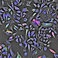
We propose a simple and compact microscope combining phase imaging with multi-color fluorescence using a standard bright-field objective. The phase image of the sample is reconstructed from a single, approximately 100 μm out-of-focus image taken under semi-coherent illumination, while fluorescence is recorded in-focus in epi-fluorescence geometry. The reproducible changes of the focus are achieved with specifically introduced chromatic aberration in the imaging system. This allows us to move the focal plane simply by changing the imaging wavelength. No mechanical movement of neither sample nor objective or any other part of the setup is therefore required to alternate between the imaging modality. Due to its small size and the absence of motorized components the microscope can easily be used inside a standard biological incubator and allows long-term imaging of cell culture in physiological conditions. A field-of-view of 1.2 mm2 allows simultaneous observation of thousands of cells with micro-meter spatial resolution in phase and multi-channel fluorescence mode. In this manuscript we characterize the system and show a time-lapse of cell culture in phase and multi-channel fluorescence recorded inside an incubator. We believe that the small dimensions, easy usage and low cost of the system make it a useful tool for biological research.
中文翻译:

使用基于色差的令人惊讶的简单显微镜进行相位和荧光成像。
我们提出了一种简单紧凑的显微镜,使用标准明场物镜将相成像与多色荧光相结合。样品的相位图像是从在半相干照明下拍摄的大约100μm的离焦图像重构而来的,而荧光则以落射荧光的几何形状进行了聚焦记录。通过在成像系统中专门引入色差,可以实现焦点的可重现变化。这使我们可以简单地通过更改成像波长来移动焦平面。因此,不需要样品,物镜或装置的任何其他部分的机械运动来在成像模态之间交替。由于其体积小且没有电动组件,因此该显微镜可以轻松地在标准生物培养箱内使用,并可以在生理条件下对细胞培养物进行长期成像。1.2 mm2的视场允许在相和多通道荧光模式下以微米级的空间分辨率同时观察数千个细胞。在本手稿中,我们对系统进行了表征,并显示了细胞培养在一段时间内的时移和在培养箱内记录的多通道荧光。我们相信该系统的小尺寸,易于使用和低成本使其成为生物学研究的有用工具。2 mm2可以在相位和多通道荧光模式下以微米级的空间分辨率同时观察数千个细胞。在本手稿中,我们对系统进行了表征,并显示了细胞培养在一段时间内的时移和在培养箱内记录的多通道荧光。我们相信该系统的小尺寸,易于使用和低成本使其成为生物学研究的有用工具。2 mm2可以在相位和多通道荧光模式下以微米级的空间分辨率同时观察数千个细胞。在本手稿中,我们对系统进行了表征,并显示了细胞培养在一段时间内的时移和在培养箱内记录的多通道荧光。我们相信该系统的小尺寸,易于使用和低成本使其成为生物学研究的有用工具。
更新日期:2020-01-17
中文翻译:
使用基于色差的令人惊讶的简单显微镜进行相位和荧光成像。
我们提出了一种简单紧凑的显微镜,使用标准明场物镜将相成像与多色荧光相结合。样品的相位图像是从在半相干照明下拍摄的大约100μm的离焦图像重构而来的,而荧光则以落射荧光的几何形状进行了聚焦记录。通过在成像系统中专门引入色差,可以实现焦点的可重现变化。这使我们可以简单地通过更改成像波长来移动焦平面。因此,不需要样品,物镜或装置的任何其他部分的机械运动来在成像模态之间交替。由于其体积小且没有电动组件,因此该显微镜可以轻松地在标准生物培养箱内使用,并可以在生理条件下对细胞培养物进行长期成像。1.2 mm2的视场允许在相和多通道荧光模式下以微米级的空间分辨率同时观察数千个细胞。在本手稿中,我们对系统进行了表征,并显示了细胞培养在一段时间内的时移和在培养箱内记录的多通道荧光。我们相信该系统的小尺寸,易于使用和低成本使其成为生物学研究的有用工具。2 mm2可以在相位和多通道荧光模式下以微米级的空间分辨率同时观察数千个细胞。在本手稿中,我们对系统进行了表征,并显示了细胞培养在一段时间内的时移和在培养箱内记录的多通道荧光。我们相信该系统的小尺寸,易于使用和低成本使其成为生物学研究的有用工具。2 mm2可以在相位和多通道荧光模式下以微米级的空间分辨率同时观察数千个细胞。在本手稿中,我们对系统进行了表征,并显示了细胞培养在一段时间内的时移和在培养箱内记录的多通道荧光。我们相信该系统的小尺寸,易于使用和低成本使其成为生物学研究的有用工具。


 京公网安备 11010802027423号