当前位置:
X-MOL 学术
›
Cytom. Part A
›
论文详情
Our official English website, www.x-mol.net, welcomes your feedback! (Note: you will need to create a separate account there.)
Cell Cycle Analysis and Relevance for Single-Cell Gating in Mass Cytometry.
Cytometry Part A ( IF 3.7 ) Pub Date : 2020-01-14 , DOI: 10.1002/cyto.a.23960 Idun D Rein 1, 2 , Heidi Ø Notø 1 , Monica Bostad 1 , Kanutte Huse 1, 3, 4 , Trond Stokke 1, 5
Cytometry Part A ( IF 3.7 ) Pub Date : 2020-01-14 , DOI: 10.1002/cyto.a.23960 Idun D Rein 1, 2 , Heidi Ø Notø 1 , Monica Bostad 1 , Kanutte Huse 1, 3, 4 , Trond Stokke 1, 5
Affiliation
|
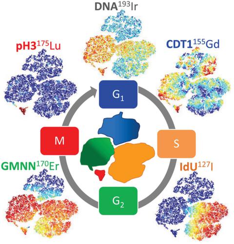
Cell cycle analysis by mass cytometry (MC) is hampered by the poor resolution of the Iridium‐labeled DNA intercalator compared to DNA‐specific fluorescent dyes. We report here a minimum cell cycle panel for MC consisting of Ir‐intercalator (DNA content), IdU (S phase), anti‐pS28HistoneH3 (mitosis), anti‐CDT1 (G1 phase) and anti‐Geminin (non‐G1 phases). Cell cycle distributions obtained by MC were not significantly different from fluorescence flow cytometry results (r 2 = 0.98, P < 0.001). Further subdivision of the G1 and G2 phases could be done with anti‐pS780RB1 (late G1) and anti‐PLK1 (late G2), respectively. A disadvantage of MC is that aggregates of cells cannot easily be removed while retaining all single cells. We have developed an analysis pipeline including unsupervised clustering by FlowSOM and subsequent single‐cell gating. When performed on cells stained with the cell cycle panel, this analysis pipeline successfully identified debris, dead/apoptotic cells, nonsingle‐cell populations and the major cell cycle phases. The presented cell cycle panel and analysis pipeline thus achieves true single‐cell analysis at the same time as any additional channels in the panel are open for phenotyping and cell cycle‐resolved expression or modification analysis. © 2020 The Authors. Cytometry Part A published by Wiley Periodicals LLC. on behalf of International Society for Advancement of Cytometry.
中文翻译:

细胞周期分析和大规模细胞计数中单细胞门控的相关性。
与 DNA 特异性荧光染料相比,铱标记的 DNA 嵌入剂的分辨率较差,这阻碍了通过质谱法 (MC) 进行的细胞周期分析。我们在此报告了 MC 的最小细胞周期面板,包括 Ir 嵌入剂(DNA 含量)、IdU(S 期)、抗 pS28HistoneH3(有丝分裂)、抗 CDT1(G 1期)和抗 Geminin(非 G 1阶段)。MC获得的细胞周期分布与荧光流式细胞术结果没有显着差异(r 2 = 0.98,P < 0.001)。G 1和 G 2阶段的进一步细分可以用抗 pS780RB1(晚期 G 1)和抗 PLK1(晚期 G 2),分别。MC 的一个缺点是在保留所有单个细胞的同时不能轻易去除细胞的聚集体。我们开发了一个分析管道,包括 FlowSOM 的无监督聚类和随后的单细胞门控。当对细胞周期面板染色的细胞进行分析时,该分析流程成功识别了碎片、死亡/凋亡细胞、非单细胞群和主要细胞周期阶段。因此,所呈现的细胞周期面板和分析管道实现了真正的单细胞分析,同时面板中的任何其他通道都对表型分析和细胞周期解析表达或修饰分析开放。© 2020 作者。由 Wiley Periodicals LLC 出版的Cytometry Part A。代表国际细胞计量学促进会。
更新日期:2020-01-14
中文翻译:
细胞周期分析和大规模细胞计数中单细胞门控的相关性。
与 DNA 特异性荧光染料相比,铱标记的 DNA 嵌入剂的分辨率较差,这阻碍了通过质谱法 (MC) 进行的细胞周期分析。我们在此报告了 MC 的最小细胞周期面板,包括 Ir 嵌入剂(DNA 含量)、IdU(S 期)、抗 pS28HistoneH3(有丝分裂)、抗 CDT1(G 1期)和抗 Geminin(非 G 1阶段)。MC获得的细胞周期分布与荧光流式细胞术结果没有显着差异(r 2 = 0.98,P < 0.001)。G 1和 G 2阶段的进一步细分可以用抗 pS780RB1(晚期 G 1)和抗 PLK1(晚期 G 2),分别。MC 的一个缺点是在保留所有单个细胞的同时不能轻易去除细胞的聚集体。我们开发了一个分析管道,包括 FlowSOM 的无监督聚类和随后的单细胞门控。当对细胞周期面板染色的细胞进行分析时,该分析流程成功识别了碎片、死亡/凋亡细胞、非单细胞群和主要细胞周期阶段。因此,所呈现的细胞周期面板和分析管道实现了真正的单细胞分析,同时面板中的任何其他通道都对表型分析和细胞周期解析表达或修饰分析开放。© 2020 作者。由 Wiley Periodicals LLC 出版的Cytometry Part A。代表国际细胞计量学促进会。


 京公网安备 11010802027423号