Nature Methods ( IF 48.0 ) Pub Date : 2020-01-13 , DOI: 10.1038/s41592-019-0688-0 Klaus C Gwosch 1 , Jasmin K Pape 1 , Francisco Balzarotti 1 , Philipp Hoess 2 , Jan Ellenberg 2 , Jonas Ries 2 , Stefan W Hell 1, 3
|
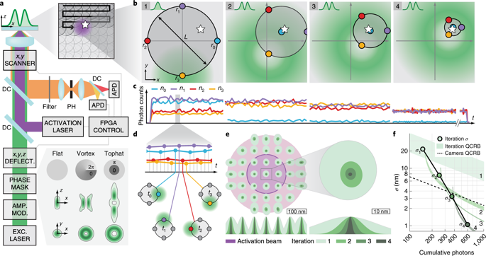
The ultimate goal of biological super-resolution fluorescence microscopy is to provide three-dimensional resolution at the size scale of a fluorescent marker. Here we show that by localizing individual switchable fluorophores with a probing donut-shaped excitation beam, MINFLUX nanoscopy can provide resolutions in the range of 1 to 3 nm for structures in fixed and living cells. This progress has been facilitated by approaching each fluorophore iteratively with the probing-donut minimum, making the resolution essentially uniform and isotropic over scalable fields of view. MINFLUX imaging of nuclear pore complexes of a mammalian cell shows that this true nanometer-scale resolution is obtained in three dimensions and in two color channels. Relying on fewer detected photons than standard camera-based localization, MINFLUX nanoscopy is poised to open a new chapter in the imaging of protein complexes and distributions in fixed and living cells.
中文翻译:
MINFLUX 纳米显微镜在细胞中提供 3D 多色纳米分辨率
生物超分辨率荧光显微镜的最终目标是提供荧光标记尺寸尺度的三维分辨率。在这里,我们展示了通过使用探测甜甜圈形激发光束定位单个可切换荧光团,MINFLUX 纳米显微镜可以为固定细胞和活细胞中的结构提供 1 至 3 nm 范围内的分辨率。通过使用探测甜甜圈最小值迭代地接近每个荧光团促进了这一进展,使分辨率在可扩展的视野中基本均匀和各向同性。哺乳动物细胞核孔复合物的 MINFLUX 成像表明,这种真正的纳米级分辨率是在三个维度和两个颜色通道中获得的。依赖于比标准的基于相机的定位更少的检测到的光子,


 京公网安备 11010802027423号