当前位置:
X-MOL 学术
›
Commun. Biol.
›
论文详情
Our official English website, www.x-mol.net, welcomes your feedback! (Note: you will need to create a separate account there.)
Hyperglycemia compromises Rat Cortical Bone by Increasing Osteocyte Lacunar Density and Decreasing Vascular Canal Volume.
Communications Biology ( IF 5.9 ) Pub Date : 2020-01-09 , DOI: 10.1038/s42003-019-0747-1 Birol Ay 1 , Kushagra Parolia 2 , Robert S Liddell 3 , Yusheng Qiu 4 , Giovanni Grasselli 4 , David M L Cooper 2 , John E Davies 1, 3
Communications Biology ( IF 5.9 ) Pub Date : 2020-01-09 , DOI: 10.1038/s42003-019-0747-1 Birol Ay 1 , Kushagra Parolia 2 , Robert S Liddell 3 , Yusheng Qiu 4 , Giovanni Grasselli 4 , David M L Cooper 2 , John E Davies 1, 3
Affiliation
|
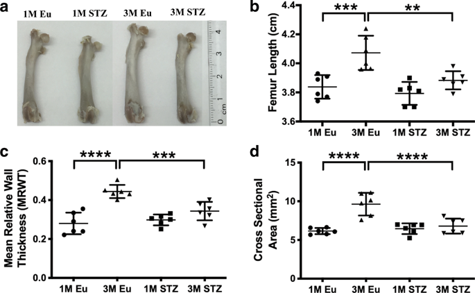
Uncontrolled diabetes is associated with increased risk of bony fractures. However, the mechanisms have yet to be understood. Using high-resolution synchrotron micro-CT, we calculated the changes in the microstructure of femoral cortices of streptozotocin-induced hyperglycemic (STZ) Wistar Albino rats and tested the mechanical properties of the mineralized matrix by nanoindentation. Total lacunar volume of femoral cortices increased in STZ group due to a 9% increase in lacunar density. However, total vascular canal volume decreased in STZ group due to a remarkable decrease in vascular canal diameter (7 ± 0.3 vs. 8.5 ± 0.4 µm). Osteocytic territorial matrix volume was less in the STZ group (14,908 ± 689 µm3) compared with healthy controls (16,367 ± 391 µm3). In conclusion, hyperglycemia increased cellularity and lacunar density, decreased osteocyte territorial matrix, and reduced vascular girth, in addition to decreasing matrix mechanical properties in the STZ group when compared with euglycemic controls.
中文翻译:

高血糖症通过增加骨细胞腔隙密度和减少血管容积来损害大鼠皮质骨。
不受控制的糖尿病会增加骨骨折的风险。但是,机制尚待了解。我们使用高分辨率同步加速器微CT,计算了链脲佐菌素诱导的高血糖(STZ)Wistar Albino大鼠股骨皮质微观结构的变化,并通过纳米压痕测试了矿化基质的力学性能。STZ组股骨皮质的腔隙总体积增加,这是由于腔隙密度增加了9%。但是,STZ组的总血管体积减少是由于血管直径显着减小(7±0.3 vs. 8.5±0.4 µm)。与健康对照组(16,367±391 µm3)相比,STZ组的骨细胞领土基质体积较小(14,908±689 µm3)。总之,高血糖会增加细胞数量和腔隙密度,
更新日期:2020-01-09
中文翻译:
高血糖症通过增加骨细胞腔隙密度和减少血管容积来损害大鼠皮质骨。
不受控制的糖尿病会增加骨骨折的风险。但是,机制尚待了解。我们使用高分辨率同步加速器微CT,计算了链脲佐菌素诱导的高血糖(STZ)Wistar Albino大鼠股骨皮质微观结构的变化,并通过纳米压痕测试了矿化基质的力学性能。STZ组股骨皮质的腔隙总体积增加,这是由于腔隙密度增加了9%。但是,STZ组的总血管体积减少是由于血管直径显着减小(7±0.3 vs. 8.5±0.4 µm)。与健康对照组(16,367±391 µm3)相比,STZ组的骨细胞领土基质体积较小(14,908±689 µm3)。总之,高血糖会增加细胞数量和腔隙密度,


 京公网安备 11010802027423号