当前位置:
X-MOL 学术
›
Ecotox. Environ. Saf.
›
论文详情
Our official English website, www.x-mol.net, welcomes your feedback! (Note: you will need to create a separate account there.)
Long-term exposure to copper induces autophagy and apoptosis through oxidative stress in rat kidneys.
Ecotoxicology and Environmental Safety ( IF 6.8 ) Pub Date : 2020-01-06 , DOI: 10.1016/j.ecoenv.2019.110158 Fang Wan 1 , Gaolong Zhong 1 , Zhijun Ning 1 , Jianzhao Liao 1 , Wenlan Yu 1 , Congcong Wang 1 , Qingyue Han 1 , Ying Li 1 , Jiaqiang Pan 1 , Zhaoxin Tang 1 , Riming Huang 2 , Lianmei Hu 1
Ecotoxicology and Environmental Safety ( IF 6.8 ) Pub Date : 2020-01-06 , DOI: 10.1016/j.ecoenv.2019.110158 Fang Wan 1 , Gaolong Zhong 1 , Zhijun Ning 1 , Jianzhao Liao 1 , Wenlan Yu 1 , Congcong Wang 1 , Qingyue Han 1 , Ying Li 1 , Jiaqiang Pan 1 , Zhaoxin Tang 1 , Riming Huang 2 , Lianmei Hu 1
Affiliation
|
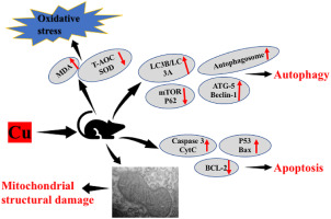
Copper (Cu) is an essential trace element for most organisms. However, excessive Cu can be highly toxic. The purpose of this study was to elucidate the mechanism underlying Cu toxicity in the kidneys of rats after treatment with CuCl2 (15 [control], 30, 60, or 120 mg/kg in the diet) for 180 days. Histological and ultrastructural changes, antioxidant enzyme activity, and the mRNA and protein levels of apoptosis and autophagy-related genes were measured. The results showed that Cu exposure led to significant accumulation of copper in kidneys and disorganized kidney morphology. The activities of total anti-oxidation capacity (T-AOC) and superoxide dismutase (SOD) in the kidneys decreased significantly, while the malondialdehyde (MDA) content increased. Furthermore, excessive Cu markedly upregulated the expression of autophagy and apoptosis-related genes (LC3A, LC3B, ATG-5, Beclin-1, Caspase3, CytC, P53, Bax), but downregulated the expression of P62, mTOR and BCL-2. Moreover, the LC3B/LC3A, ATG-5, Beclin-1, P53, Caspase3 proteins were up-regulated while P62 was down-regulated in the kidney tissues of the treatment groups. Overall, these findings provide strong evidence that excess Cu can trigger autophagy and apoptosis via the mitochondrial pathway by inducing oxidative stress in rat kidneys.
中文翻译:

长期暴露于铜会通过大鼠肾脏的氧化应激诱导自噬和凋亡。
铜(Cu)是大多数生物必不可少的微量元素。但是,过量的铜可能具有剧毒。这项研究的目的是阐明用CuCl2(饮食中的15 [对照组],30、60或120 mg / kg)治疗180天后,大鼠肾脏中Cu毒性的潜在机制。测量组织学和超微结构变化,抗氧化酶活性以及凋亡和自噬相关基因的mRNA和蛋白水平。结果表明,铜暴露导致肾脏中大量铜积累和肾脏形态紊乱。肾脏中总抗氧化能力(T-AOC)和超氧化物歧化酶(SOD)的活性显着降低,而丙二醛(MDA)含量增加。此外,过量的铜明显上调自噬和凋亡相关基因(LC3A,LC3B,ATG-5,Beclin-1,Caspase3,CytC,P53,Bax)的表达,但下调P62,mTOR和BCL-2的表达。此外,在治疗组的肾脏组织中,LC3B / LC3A,ATG-5,Beclin-1,P53,Caspase3蛋白被上调,而P62被下调。总体而言,这些发现提供了有力的证据,表明过量的铜可通过诱导大鼠肾脏的氧化应激,通过线粒体途径触发自噬和凋亡。
更新日期:2020-01-07
中文翻译:
长期暴露于铜会通过大鼠肾脏的氧化应激诱导自噬和凋亡。
铜(Cu)是大多数生物必不可少的微量元素。但是,过量的铜可能具有剧毒。这项研究的目的是阐明用CuCl2(饮食中的15 [对照组],30、60或120 mg / kg)治疗180天后,大鼠肾脏中Cu毒性的潜在机制。测量组织学和超微结构变化,抗氧化酶活性以及凋亡和自噬相关基因的mRNA和蛋白水平。结果表明,铜暴露导致肾脏中大量铜积累和肾脏形态紊乱。肾脏中总抗氧化能力(T-AOC)和超氧化物歧化酶(SOD)的活性显着降低,而丙二醛(MDA)含量增加。此外,过量的铜明显上调自噬和凋亡相关基因(LC3A,LC3B,ATG-5,Beclin-1,Caspase3,CytC,P53,Bax)的表达,但下调P62,mTOR和BCL-2的表达。此外,在治疗组的肾脏组织中,LC3B / LC3A,ATG-5,Beclin-1,P53,Caspase3蛋白被上调,而P62被下调。总体而言,这些发现提供了有力的证据,表明过量的铜可通过诱导大鼠肾脏的氧化应激,通过线粒体途径触发自噬和凋亡。


 京公网安备 11010802027423号