当前位置:
X-MOL 学术
›
Cell Death Dis.
›
论文详情
Our official English website, www.x-mol.net, welcomes your feedback! (Note: you will need to create a separate account there.)
Epigenetic priming by Dot1l in lymphatic endothelial progenitors ensures normal lymphatic development and function.
Cell Death & Disease ( IF 9 ) Pub Date : 2020-01-06 , DOI: 10.1038/s41419-019-2201-1 Hyunjin Yoo 1 , Young Jae Lee 2 , Chanhyeok Park 1 , Dabin Son 1 , Dong Yoon Choi 1 , Ji-Hyun Park 1 , Hee-Jin Choi 1 , Hyun Woo La 1 , Yun-Jung Choi 1 , Eun-Hye Moon 2 , Dieter Saur 3, 4 , Hyung Min Chung 5 , Hyuk Song 1 , Jeong Tae Do 1 , Hoon Jang 6 , Dong Ryul Lee 6 , Chankyu Park 1 , Ok-Hee Lee 6 , Ssang-Goo Cho 1 , Seok-Ho Hong 7 , Gu Kong 8 , Jin-Hoi Kim 1 , Youngsok Choi 1 , Kwonho Hong 1
Cell Death & Disease ( IF 9 ) Pub Date : 2020-01-06 , DOI: 10.1038/s41419-019-2201-1 Hyunjin Yoo 1 , Young Jae Lee 2 , Chanhyeok Park 1 , Dabin Son 1 , Dong Yoon Choi 1 , Ji-Hyun Park 1 , Hee-Jin Choi 1 , Hyun Woo La 1 , Yun-Jung Choi 1 , Eun-Hye Moon 2 , Dieter Saur 3, 4 , Hyung Min Chung 5 , Hyuk Song 1 , Jeong Tae Do 1 , Hoon Jang 6 , Dong Ryul Lee 6 , Chankyu Park 1 , Ok-Hee Lee 6 , Ssang-Goo Cho 1 , Seok-Ho Hong 7 , Gu Kong 8 , Jin-Hoi Kim 1 , Youngsok Choi 1 , Kwonho Hong 1
Affiliation
|
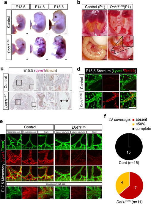
Proper functioning of the lymphatic system is required for normal immune responses, fluid balance, and lipid reabsorption. Multiple regulatory mechanisms are employed to ensure the correct formation and function of lymphatic vessels; however, the epigenetic modulators and mechanisms involved in this process are poorly understood. Here, we assess the regulatory role of mouse Dot1l, a histone H3 lysine (K) 79 (H3K79) methyltransferase, in lymphatic formation. Genetic ablation of Dot1l in Tie2(+) endothelial cells (ECs), but not in Lyve1(+) or Prox1(+) lymphatic endothelial cells (LECs) or Vav1(+) definitive hematopoietic stem cells, leads to catastrophic lymphatic anomalies, including skin edema, blood-lymphatic mixing, and underdeveloped lymphatic valves and vessels in multiple organs. Remarkably, targeted Dot1l loss in Tie2(+) ECs leads to fully penetrant lymphatic aplasia, whereas Dot1l overexpression in the same cells results in partially hyperplastic lymphatics in the mesentery. Genetic studies reveal that Dot1l functions in c-Kit(+) hemogenic ECs during mesenteric lymphatic formation. Mechanistically, inactivation of Dot1l causes a reduction of both H3K79me2 levels and the expression of genes important for LEC development and function. Thus, our study establishes that Dot1l-mediated epigenetic priming and transcriptional regulation in LEC progenitors safeguard the proper lymphatic development and functioning of lymphatic vessels.
中文翻译:

Dot111在淋巴管内皮祖细胞中进行表观遗传启动可确保正常的淋巴发育和功能。
正常的免疫反应,体液平衡和脂质重吸收需要淋巴系统正常运转。采用多种调节机制来确保淋巴管的正确形成和功能;然而,对此过程涉及的表观遗传调节剂和机制了解甚少。在这里,我们评估小鼠Dot11,组蛋白H3赖氨酸(K)79(H3K79)甲基转移酶在淋巴形成中的调节作用。在Tie2(+)内皮细胞(EC)中进行Dot1l的遗传消融,但在Lyve1(+)或Prox1(+)淋巴管内皮细胞(LEC)或Vav1(+)最终造血干细胞中未进行遗传切除,导致灾难性淋巴异常,包括皮肤水肿,血液淋巴混合以及多器官中不发达的淋巴瓣膜和血管。值得注意的是 在Tie2(+)EC中有针对性的Dot11丢失会导致完全渗透性淋巴发育不良,而同一细胞中Dot11的过表达会导致肠系膜部分增生的淋巴结。遗传学研究表明,在肠系膜淋巴结形成过程中,Dot11在c-Kit(+)造血EC中起作用。从机制上讲,Dot11的失活会导致H3K79me2水平的降低以及对LEC发育和功能很重要的基因的表达降低。因此,我们的研究建立了LEC祖细胞中Dot11介导的表观遗传启动和转录调控可维护淋巴管的正常淋巴发育和功能。遗传学研究表明,在肠系膜淋巴结形成过程中,Dot11在c-Kit(+)造血EC中起作用。从机制上讲,Dot11的失活会导致H3K79me2水平的降低以及对LEC发育和功能很重要的基因的表达降低。因此,我们的研究建立了LEC祖细胞中Dot11介导的表观遗传启动和转录调控可维护淋巴管的正常淋巴发育和功能。遗传学研究表明,在肠系膜淋巴结形成过程中,Dot11在c-Kit(+)造血EC中起作用。从机理上讲,Dot11的失活导致H3K79me2水平的降低以及对LEC发育和功能重要的基因的表达降低。因此,我们的研究建立了LEC祖细胞中Dot11介导的表观遗传启动和转录调控可维护淋巴管的正常淋巴发育和功能。
更新日期:2020-01-06
中文翻译:
Dot111在淋巴管内皮祖细胞中进行表观遗传启动可确保正常的淋巴发育和功能。
正常的免疫反应,体液平衡和脂质重吸收需要淋巴系统正常运转。采用多种调节机制来确保淋巴管的正确形成和功能;然而,对此过程涉及的表观遗传调节剂和机制了解甚少。在这里,我们评估小鼠Dot11,组蛋白H3赖氨酸(K)79(H3K79)甲基转移酶在淋巴形成中的调节作用。在Tie2(+)内皮细胞(EC)中进行Dot1l的遗传消融,但在Lyve1(+)或Prox1(+)淋巴管内皮细胞(LEC)或Vav1(+)最终造血干细胞中未进行遗传切除,导致灾难性淋巴异常,包括皮肤水肿,血液淋巴混合以及多器官中不发达的淋巴瓣膜和血管。值得注意的是 在Tie2(+)EC中有针对性的Dot11丢失会导致完全渗透性淋巴发育不良,而同一细胞中Dot11的过表达会导致肠系膜部分增生的淋巴结。遗传学研究表明,在肠系膜淋巴结形成过程中,Dot11在c-Kit(+)造血EC中起作用。从机制上讲,Dot11的失活会导致H3K79me2水平的降低以及对LEC发育和功能很重要的基因的表达降低。因此,我们的研究建立了LEC祖细胞中Dot11介导的表观遗传启动和转录调控可维护淋巴管的正常淋巴发育和功能。遗传学研究表明,在肠系膜淋巴结形成过程中,Dot11在c-Kit(+)造血EC中起作用。从机制上讲,Dot11的失活会导致H3K79me2水平的降低以及对LEC发育和功能很重要的基因的表达降低。因此,我们的研究建立了LEC祖细胞中Dot11介导的表观遗传启动和转录调控可维护淋巴管的正常淋巴发育和功能。遗传学研究表明,在肠系膜淋巴结形成过程中,Dot11在c-Kit(+)造血EC中起作用。从机理上讲,Dot11的失活导致H3K79me2水平的降低以及对LEC发育和功能重要的基因的表达降低。因此,我们的研究建立了LEC祖细胞中Dot11介导的表观遗传启动和转录调控可维护淋巴管的正常淋巴发育和功能。


 京公网安备 11010802027423号